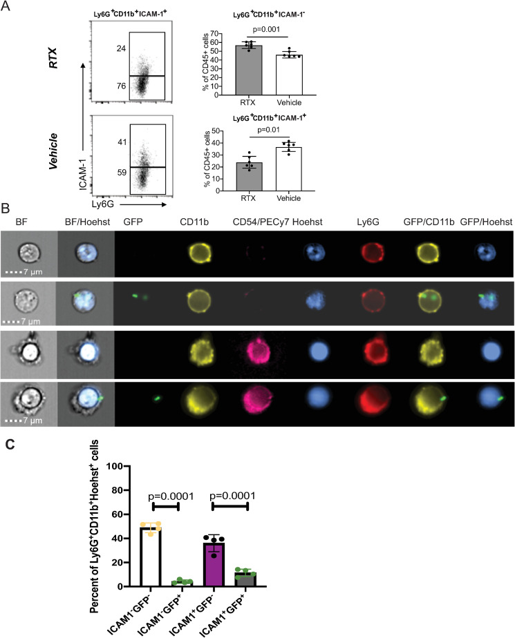
Fig 6. RTX treatment reduces the frequencies of CD11b+ Ly6G+ICAM-1+ myeloid cells in the infected corneas.
A. Histogram plots show gating and frequencies of CD11b+ Ly6G+ICAM-1- and CD11b+Ly6G+ ICAM-1+ cells. Data represent 6 infected corneas (1 per mouse) per condition per cohort. The experiment was repeated twice. Bar graphs show mean values; symbols represent individual mouse. P-values by Student’s t-test. B. Representative ImageStream images of CD11b+ Ly6G+ICAM-1+ and CD11b+ Ly6G+ ICAM-1- cells. Mice were infected with 5×10⁵ CFU of GFP-expressing P. aeruginosa 6294 (Green) per cornea; tissue was harvested at 24h post-challenge for analysis. Cellular suspensions were prepared with collagenase digestion and stained for CD45, CD11b, Ly6G, ICAM-1, and DNA (Hoechst) to identify myeloid infiltrates. CD11b+ Ly6G+ cells show banded or multi-lobed nuclear morphology typical of neutrophils. C. Quantification of CD11b+ Ly6G+ICAM-1+ and CD11b+ Ly6G+ ICAM-1- myeloid cells containing intracellular P. aeruginosa-GFP (p-values by one-way ANOVA). Cumulatively, data show that nociceptor presence alters myeloid phenotypes in the infected corneas, enriching for CD11b+ ICAM-1+ Ly6G+ myeloid cells in vehicle-treated mice when compared to RTX-treated mice.

