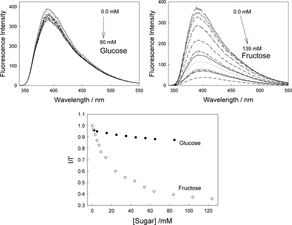
Figure 15.9.
Emission spectra of the CSTBA doped contact lens, pH 8.0 buffer / methanol (2:1), with increasing concentrations of glucose (top left) and corresponding spectra with increasing concentrations of fructose (top right). λex = 320 nm. Intensity ratio plot for CSTBA doped contact lens towards both glucose and fructose (bottom), where I and I’ are the intensities in the presence and absence of sugar respectively at λem max.

