. 2021 Apr 15;10:e67552. doi: 10.7554/eLife.67552

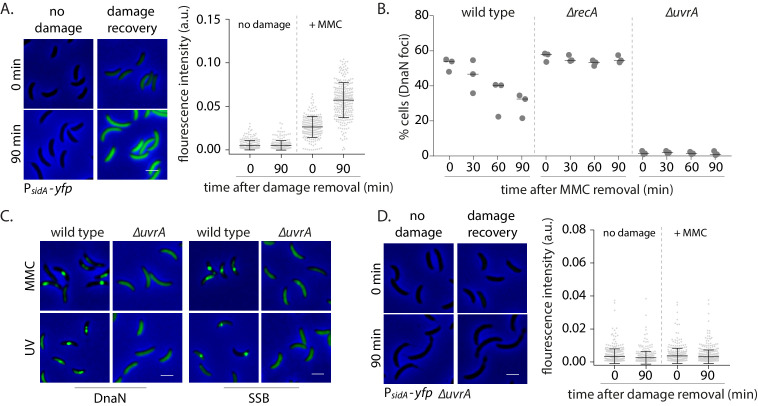
Figure 3. Nucleotide excision repair (NER) generates long ssDNA gaps for localization of replisome components in non-replicating cells.

(A) SOS induction was measured by assessing the expression of yfp from an SOS-inducible promoter (PsidA-yfp). On the left are representative images of cells expressing the reporter at 0 or 90 min after MMC removal and control cells (no damage). On the right, total fluorescence intensity normalized to cell area is plotted for both time points for cells with or without damage treatment. Each dot represents a single cell. Mean and SD are shown in black (n ≥ 219). (B) Percentage wild type, ∆recA, or ∆uvrA swarmer cells with DnaN foci at 0, 30, 60, and 90 min after DNA damage recovery (n ≥ 308 cells, three independent repeats). (C) Representative images of wild type or ∆uvrA swarmer cells with SSB-YFP or DnaN-YFP, treated with MMC or UV. (D) As (A) for cells lacking uvrA (n ≥ 325).

Figure 3—source data 1. Source data related to panels in Figure 3.

Figure 3.

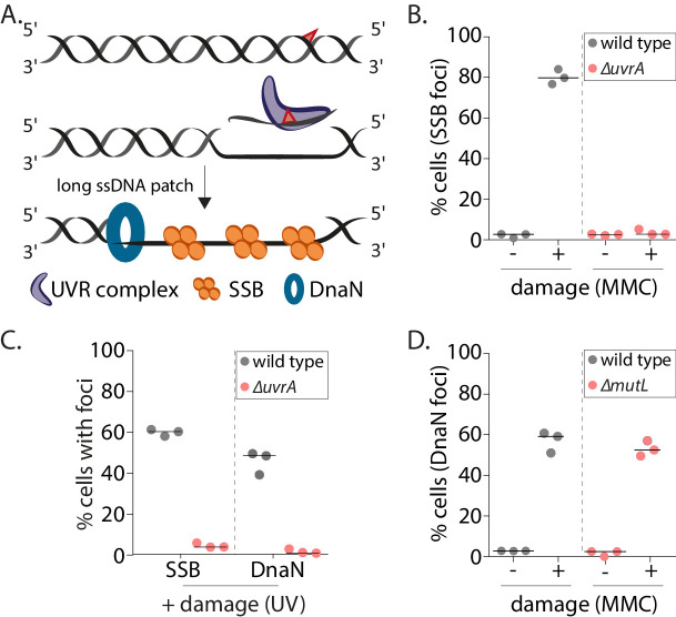
Figure 3—figure supplement 1. Nucleotide excision repair (NER) generates long ssDNA gaps for localization of replisome components in non-replicating cells.

Figure 3—figure supplement 1.

(A) Schematic of mechanism of long ssDNA gap generation by nucleotide excision repair (NER). (B) Percentage wild type or ∆uvrA swarmer cells with SSB foci with (+MMC) or without (-, control) damage treatment (n ≥ 325 cells, three independent repeats, wild type data from Figure 2B). (C) Percentage wild type or ∆uvrA swarmer cells with DnaN or SSB foci after DNA damage (UV) (n ≥ 340 cells, three independent repeats). (D) Percentage wild type or ∆mutL swarmer cells with DnaN foci with (+MMC) or without (-, control) damage treatment (n ≥ 324 cells, three independent repeats, wild type data from Figure 2B). MMC: mitomycin C.
Figure 3—figure supplement 1—source data 1. Source data related to panels in Figure 3—figure supplement 1.