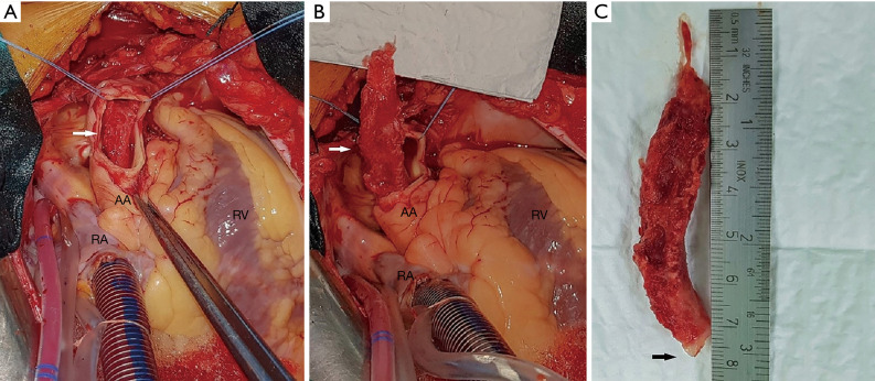
Figure 2.
Surgical view of the intra-aortic, partial and wholly removed thrombus mass. (A) Intra-aortic presentation of the thrombus through the transverse aortotomy. (B) Distal part of thrombus extirpated from the aortic arch. (C) Completely extracted 80 mm × 13 mm thrombus mass. White arrow, thrombus mass; black arrow, pedicle of thrombus. LV, left ventricle; RA, right atrium; AA, ascending aorta.

