Figure 2.
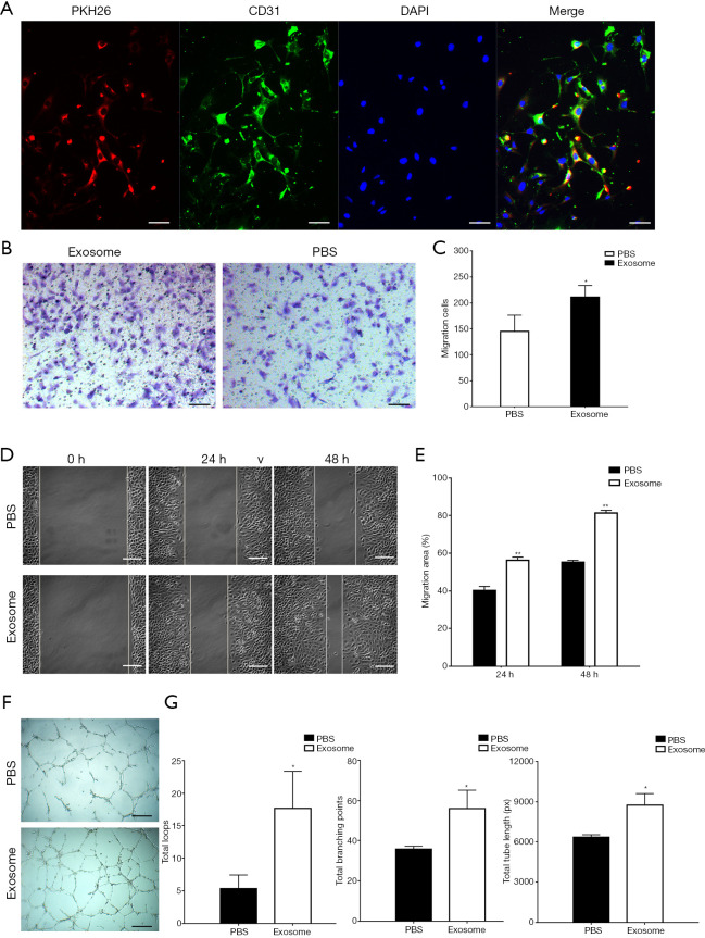
The hAFMSCExos promoted migration and tube formation of HUVECs after OGD in vitro. (A) HUVECs uptaken PKH26-labeled hAFMSCExo by fluorescence microscopy analysis; scale bar: 100 µm. (B,C) The migration of HUVECs stimulated by hAFMSCExos or PBS after OGD was analyzed by transwell assay; scale bar: 100 µm; *, P<0.05. (D,E) The migration of HUVECs treated with hAFMSCExos or PBS after OGD in scratch wound assay; scale bar: 50 µm; **, P<0.01. (F) The tube formation ability of treated with hAFMSCExos or PBS after OGD in capillary network formation assay; scale bar: 50 µm. (G) The total loops, total branching points, and total tube length of HUVECs treated with hAFMSCs-Exos or PBS after OGD in capillary network formation assay. *, P<0.05; hAFMSCExos, human amniotic fluid mesenchymal stem cell-derived exosomes; HUVEC, human umbilical vein endothelial cells; OGD, oxygen and glucose deprivation; PBS, phosphate-buffered saline.

