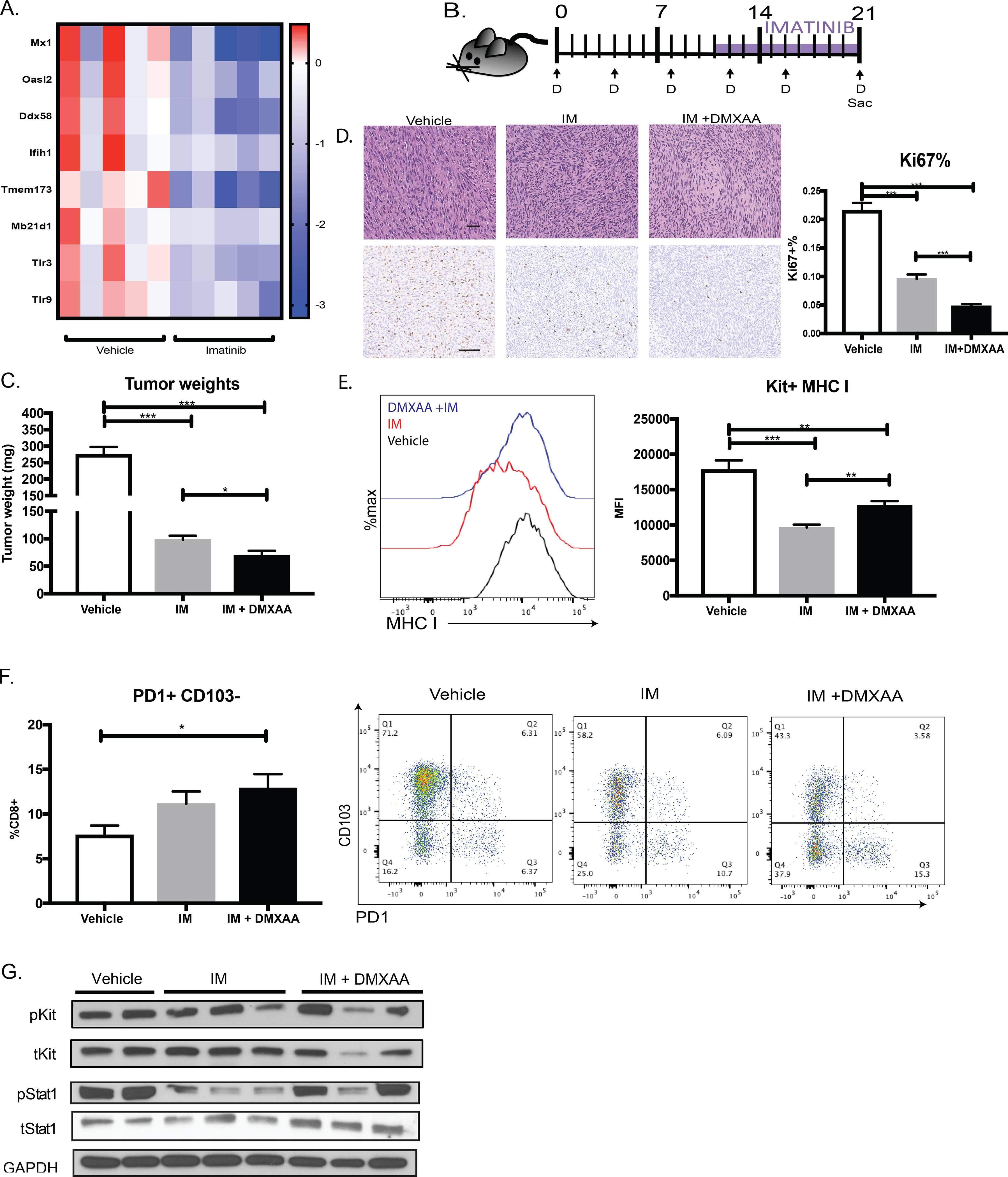
Figure 6. Stimulation of type I IFN signaling augments imatinib therapy in GIST.

(A) Gene expression of innate immune nucleic acid sensor genes from RNAseq of KitV558Δ/+ GIST treated with and without 3 weeks of imatinib. Each column represents an individual mouse. (B) KitV558Δ/+ mice were treated with DMXAA (D) for 10 days and given 11 days of imatinib. Controls received vehicle or imatinib from day 10 to day 21. (C) Tumor weights of KitV558Δ/+ GIST treated with vehicle (7.5% NaCO2), imatinib (11 days), or both. Unpaired two-sample t-test of treatment performed against vehicle controls. Data represent mean ± SEM, * p-value < 0.05, ** p-value <0.01, *** p-value <0.001. N= 4–5 mice/group, repeated twice. (D) Representative tumor histology (40x) and Ki67 staining (20x) shown, where bar measures 50μm. Percentage of Ki67+ cells calculated by stained nuclei divided by total nuclei in five 100 × 100 μm HPF of at least three different tumors per treatment group (E) MHC I expression on Kit+ tumor cells from (C) by MFI using flow cytometry analysis. (F) PD1 and CD103 expression on intratumoral CD8+ T cells in mice treated from (C). (G) Immunoblot of STAT1 and KIT signaling in GIST treated with vehicle, imatinib, or imatinib and DMXAA.
