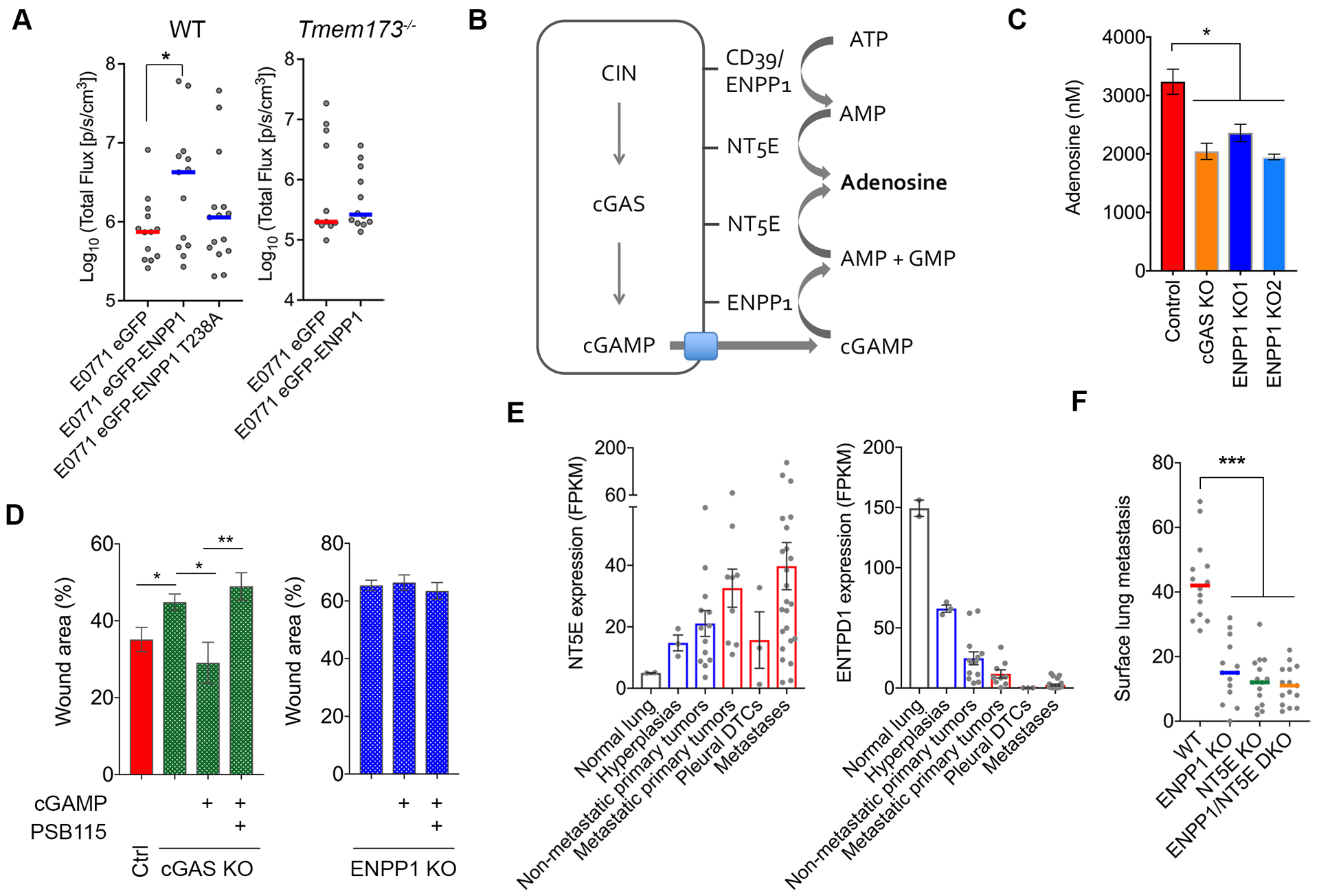
Figure 2. ENPP1 promotes extracellular adenosine production.

(A) Left, total bioluminescence imaging of WT or Tmem173−/− animals inoculated with E0771 cells expressing WT or enzymatically weakened ENPP1 (T328A), bars represent median, n = 13–15 mice per group for the WT animals and 11–12 for the Tmem173−/− animals, * p < 0.05, Welch t-test. (B) Schematic showing the generation of adenosine from extracellular cGAMP and ATP hydrolysis. (C) Normalized adenosine concentration (per 107 cells after 16 hours incubation in serum-free media) in conditioned media of control, Cgas-KO, Enpp1-KO 4T1 cells, bars represent mean ± s.e.m., n = 4 independent experiments, *p<0.05, two-sided t-test. (D) Percent wound remaining after 24 hours in control, Cgas-KO, and Enpp1-KO 4T1 cells treated with cGAMP or cGAMP and the adenosine receptor blocker, PSB115. (E) NT5E and ENTPD1 mRNA expression in various stages of lung adenocarcinoma progression, bars represent mean ± s.e.m. (F) Surface lung metastases after tail vein injection of control, Enpp1-KO, Nt5e-KO, and Enpp1/Nt5e double KO 4T1 cells, bars represent median, n = 15 animals per condition, **** p < 0.001, two-sided Mann-Whitney test.
