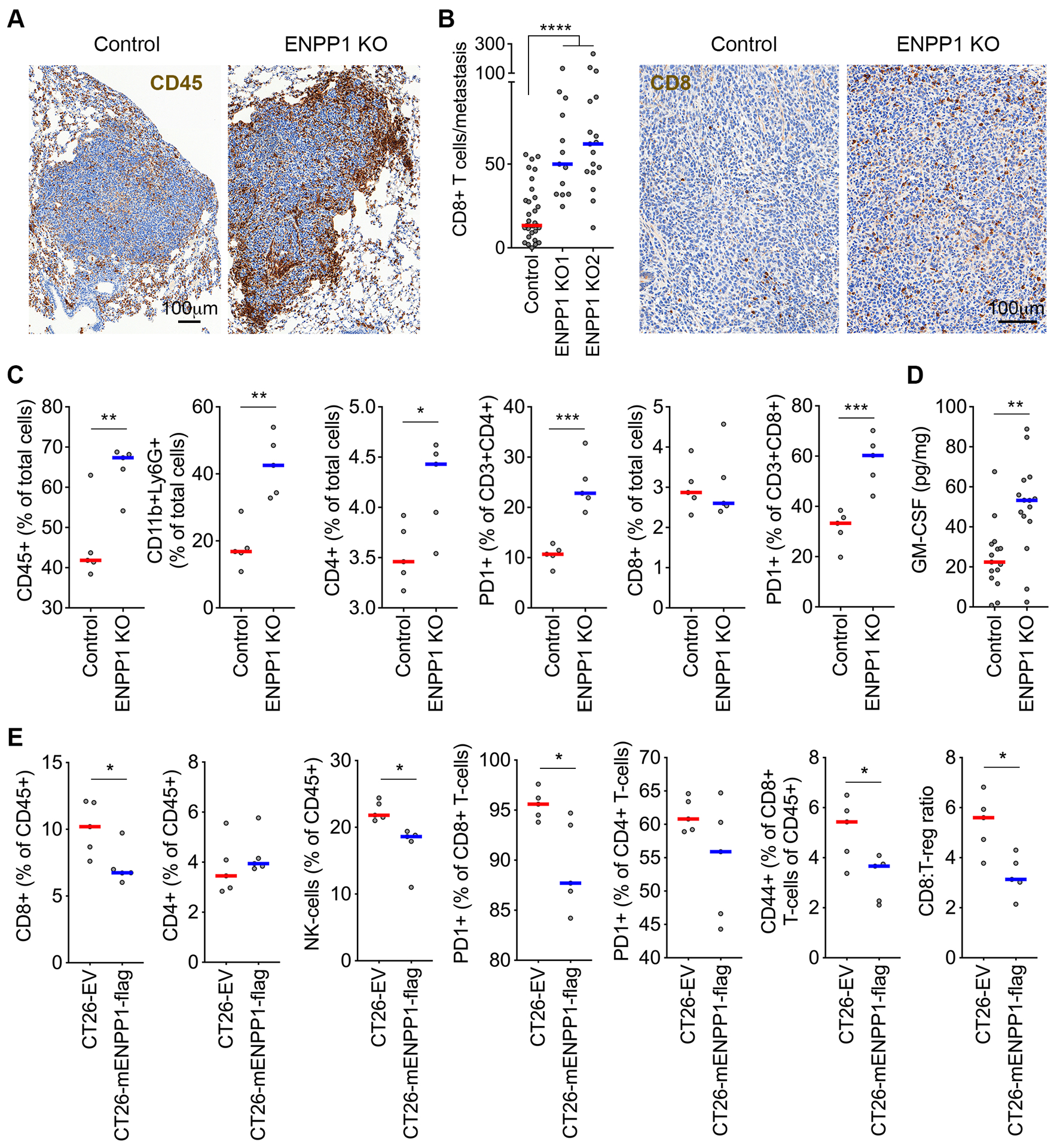
Figure 3. ENPP1 reduces tumor immune infiltration.

(A) Representative immunohistochemistry (IHC) of control and ENPP1-knockout TNBC lung metastases stained using an anti-CD45 antibody. (B) The number of metastasis-infiltrating CD8+ T-cells (left) and representative IHC of control ENPP1-knockout TNBC lung metastases stained using anti-CD8 antibody (right), bars represent median, n = 13–31 metastases, **** p<0.0001, two-sided Mann-Whitney test. (C) Percentage of CD45+, CD11b+Ly6G+, CD4+, and CD8+ cells out of the total cells as well as the percentage of PD1+ cells out of the CD3+CD4+ and CD3+CD8+ cells obtained from dissociated lungs after injection with control or ENPP1-knockout 4T1 cells, n = 5 animals per group. (D) GM-CSF levels measured in orthotopically transplanted control and ENPP1-knockout tumors, bars represent median, n = 15 tumors per condition, ** p<0.01, two-sided Mann-Whitney test. (E) Percentage of CD8+ T-cells, CD4+ T-cells (and the PD1+ and CD44+ fractions of thereof), and NK-cells obtained from dissociated subcutaneously transplanted control and ENPP1 expressing CT26 tumors, n = 5 animals per group, bars represent median, * p < 0.05.
