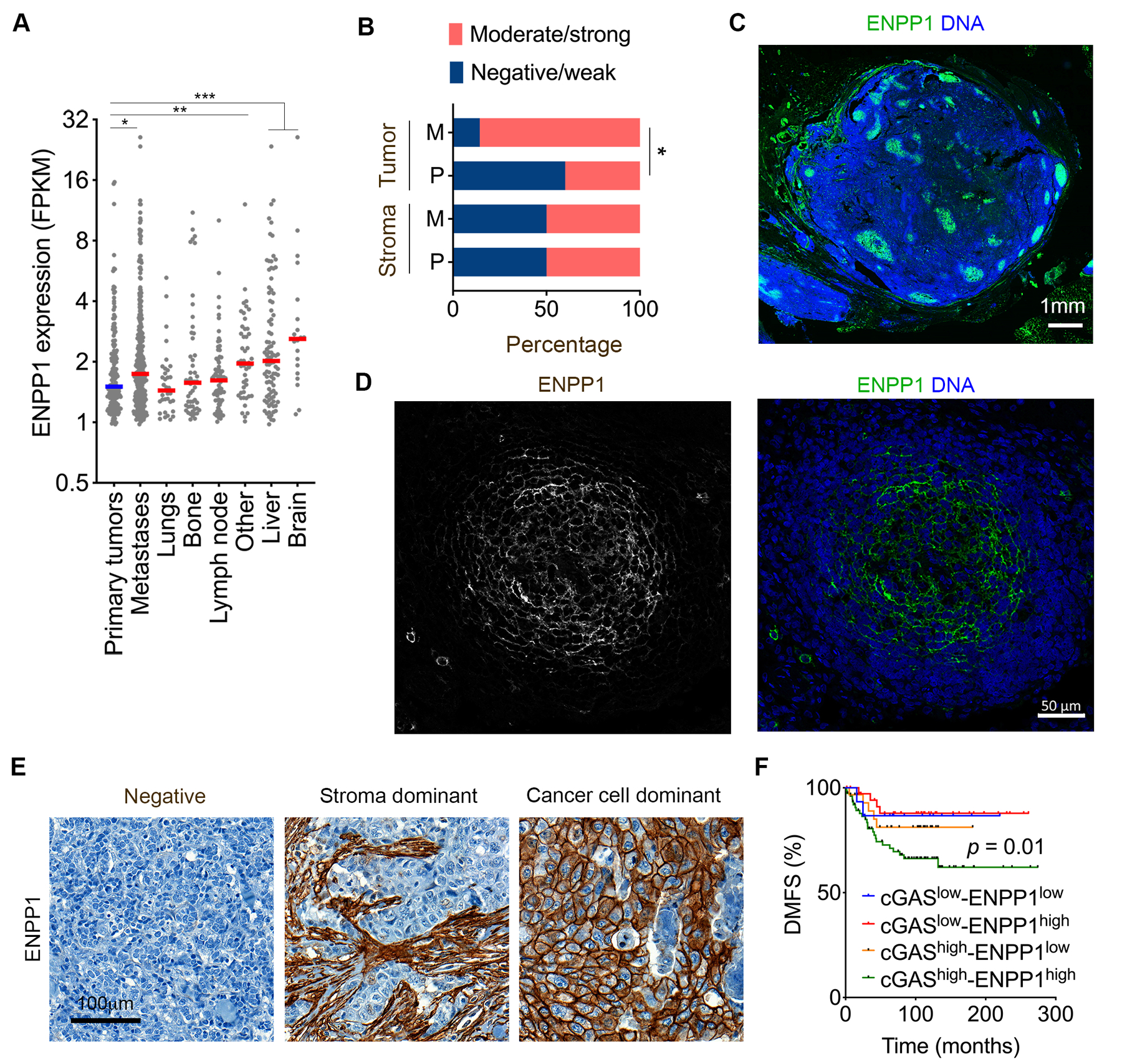
Figure 5. ENPP1 expression is associated with metastasis in human cancer.

(A) ENPP1 expression across primary and metastatic tumors, stratified by the site of metastasis, n = 180 tumors for primary tumors and 331 tumors for metastases, bars represent median, * p < 0.05, ** p < 0.01, *** p < 0.001. (B) Percentage of mucosal melanoma patients with tumor-specific or stromal specific ENPP1 staining patterns in primary as well as metastatic mucosal melanoma human tumor samples, *p < 0.05, χ2-test. (C-D) Representative immunofluorescence images of low (C) and high (D) magnification images of lymph node metastases from mucosal melanoma stained using DAPI (DNA) and anti-ENPP1 antibody showing selective membrane staining of ENPP1 on metastatic cancer cells. Scale bar 1 mm (C) and 50 μm (D). (E) Representative images of human TNBCs stained using anti-ENPP1 antibody, scale bar 100 μm. (F) Distant-metastasis-free survival in patients with TNBC stratified based on their ENPP1 and cGAS expression n = 159, significance tested using log-rank test.
