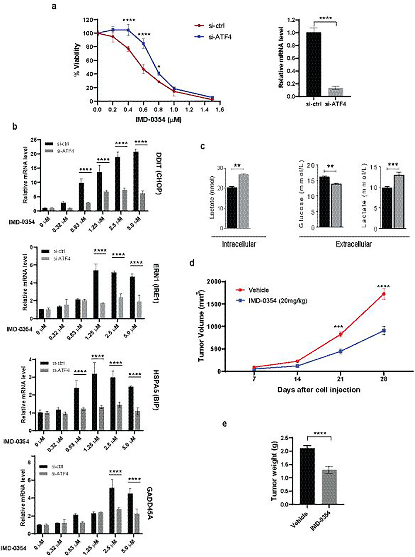
Figure 8. Characterization of IMD-0354 Effect on Melanoma in Culture and In Vivo.

(a) A375 cells were transfected with si-ctrl or si-ATF4 and treated with increasing concentrations of IMD-0354. Cell proliferation was measured after 72h by ATPlite (left). ATF4 knockdown efficiency was confirmed by qPCR (right). (b) A375 cells were transfected with si-ctrl or si-ATF4 for 48h followed by treatment with IMD-0354 for 6h at indicated concentrations. RNA was extracted and subjected to qPCR analysis for UPR-related genes. (c) IMD-0354 treatment enhanced lactate production and suppressed glucose uptake. A431 cells were treated with IMD-0354 (2 μM) for 72h. Extracellular glucose and both extracellular and intracellular lactate levels were determined by GC-MS. (d, e) SW1 cells were injected into the right flank of male C3H/HeJ mice. After tumor establishment (~150mm3), mice were treated with 20 mg/kg of IMD-0354 daily intraperitoneally. Tumor volume was monitored for three weeks (d). Tumors were weighed at the end of the experiment (e). Statistical analysis was performed by two-way ANOVA for viability assay and tumor growth. Unpaired t-test was used for the comparison of two groups. Data are shown as the mean ± SD, n = 3. *P ≤ 0.05, **P ≤ 0.01, ***P ≤ 0.001, ****P ≤ 0.0001. For animal experiment, statistical analysis was performed by two-way ANOVA for time-dependent tumor volume changes (d) and unpaired t-test was used for the comparison of end point tumor weight (e). Data are shown as the mean ± SEM, n = 8. ***P ≤ 0.001, ****P ≤ 0.0001.
