
Fig. 5. TAM-deficient microglia can neither phagocytose nor organize plaques.
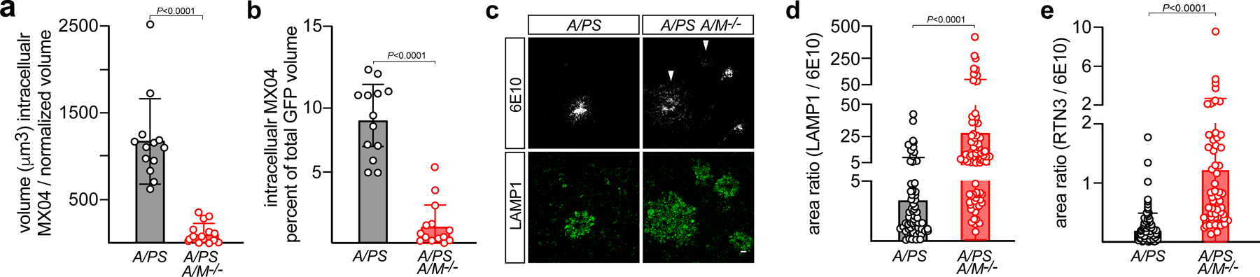
(a, b) MX04-labeled Aβ plaque material engulfed within GFP+ microglia, imaged in vivo, in 16 mo. APP/PS1 (A/PS) versus APP/PS1Axl−/−Mertk−/− (A/PS A/M−/−) cortex, normalized to imaging volume (a) and the volume of GFP+ cells (b). (c) Representative images of the halo of LAMP1+ dystrophic membranes (green, lower panels) that surround 6E10+ plaques in 12 mo APP/PS1 (A/PS; left) versus APP/PS1Axl−/−Mertk−/− (A/PS A/M−/−; right) cortex. Arrowheads mark weakly-staining, diffuse 6E10+ plaques, which are more common in the APP/PS1Axl−/−Mertk−/− brain (see also Extended Data Fig. 5). Scale bar: 10μm. (d) Quantification of the ratio of LAMP1+ area to 6E10+ plaque area across all plaque sizes, both dense-core and diffuse. (e) Quantification of ratio RTN3+ area to 6E10+ plaque area across all plaque sizes, both dense-core and diffuse. Data are 13–15 volumetric images from n= 3 and 4 mice for APP/PS1 and APP/PS1Axl−/−Mertk−/−, respectively (a–b). Data are 94–113 plaques (d) and 56–95 plaques (e) investigated from N ≥ 3 sections per mouse from n = 3 mice of each genotype. Mann-Whitney test (a, b, d, e). Data represented as mean ±1 STD.