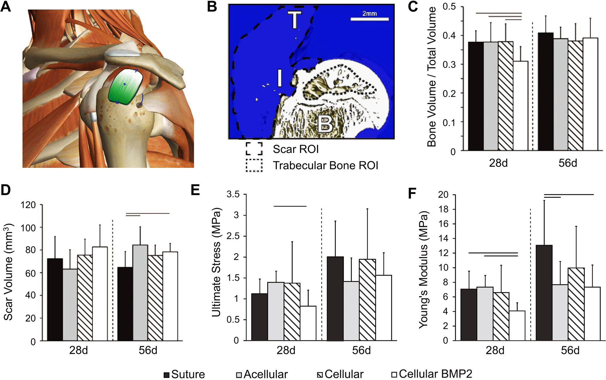
Figure 4.

In vivo evaluation of PLGA electrospun nanofiber mats with mineral gradients for tendon-to-bone repair. A) Schematic illustration showing the human shoulder patched with a mineral-graded nanofiber mat. B) 3D reconstruction of the repaired attachment, in which B, I, and T represent bone, insertion, and tendon, respectively. C) Bone volume over total volume, D) scar volume, E) ultimate stress, and F) Young’s modulus as a function of implantation time. Significance is indicated by lines over bars (p < 0.05). For all data shown in C–F), “Suture”, “Acellular”, “Cellular”, and “Cellular BMP2” indicate the group without ASCs and scaffold, the group repaired with a scaffold but without ASCs, the group repaired with ASC-seeded scaffold, and the group repaired with BMP2-transduced ASC-seeded scaffold, respectively. A–F) Adapted with permission.[66] Copyright 2015, Mary Ann Liebert, Inc.
