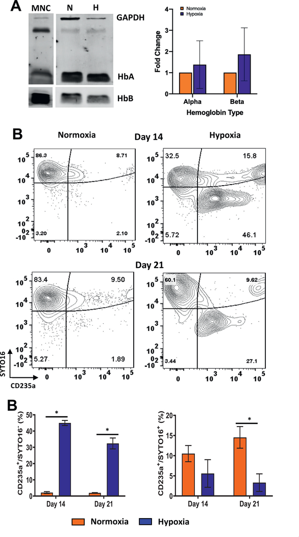
Figure 7.
Hypoxia promotes enucleation of erythroid cells. (A) Immunoblotting of α- and β-hemoglobin expression in CD34+ cell cultures incubated in normoxia or hypoxia and in cord blood-derived mononuclear cells (MNCs). Average protein expression (§SE, n = 3) normalized to GAPDH is illustrated. (B) Enucleation was determined by SYTO16 staining. Contour plots for CD235a+ and SYTO16+ cells in cultures incubated in hypoxia or normoxia. (C) Percentages of enucleated (CD235a+/SYTO16−) and nucleated (CD235a+/SYTO16+) erythroid cells in the CD34−/Live population on days 14 and 21 are represented as the mean with standard error (n = 4). Statistical analysis was performed using the Mann-Whitney test, and p values ≤ 0.05 were considered significant. *p < 0.05, **p < 0.005, ***p < 0.0005.

