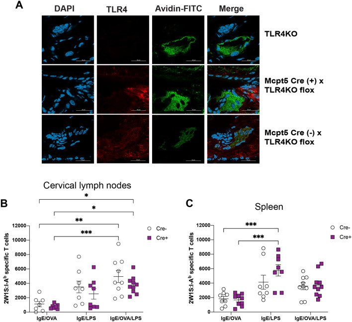
Figure 5.
TLR4 expression on mast cells is not entirely responsible for the IgE/OVA- and LPS-mediated expansion of antigen-specific CD4 + T cells. (A) Tissue ear sections from TLR4-deficient mice (top), Mcpt5 Cre ( −) x Tlr4KO flox (middle), and Mcpt Cre ( +) x Tlr4KO flox (bottom) were imaged using confocal microscopy (see “Materials and methods” section). Red (PE) indicates TLR4 stain, green (FITC) indicates MC-granule-specific avidin stain, and DAPI (blue) indicates cell nuclei. Image shows that Cre ( +) MCs lack TLR4 expression in comparison to Cre ( −) MCs. (B,C) Mast cell-specific TLR4-deficient mice (Cre +) and littermate controls (Cre −) were immunized intradermally in the ears with 2W1S peptide and the indicated combination of IgE, OVA, and LPS. Cervical draining lymph nodes (B) and spleens (C) were analyzed 10 days later for the numbers of 2W1S-specific CD4+ T cells. Results are cumulative data from 3 independent experiments with n = 7–10 mice total and shown as mean ± SEM. Scale bar, 25 µm. *P < 0.05 **P < 0.01, ***P < 0.001, **** P < 0.0001. Statistical analysis was performed using one-way ANOVA with Tukey’s multiple comparisons test.

