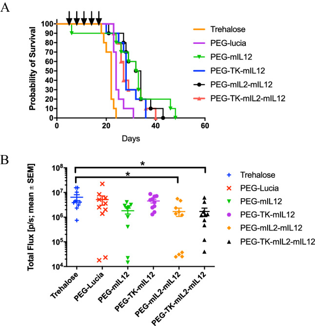
Figure 5.

Activity of l-PEI/DNA nanoparticles in a B16F10 syngeneic, metastatic model of cancer in the lungs in C57BL/6 mice represented by a Kaplan Meier survival plot. Mice were inoculated intravenously via the tail vein with B16F10-Red-FLuc murine melanoma cells and treated with nanoparticles at 3-d intervals, beginning at Day 5 (post tumor cell inoculation) as indicated by the arrows above the plot. Nanoparticle formulations of plasmids PEG-mIL12 (green line, filled upturned triangle), PEG-TK-mIL12, PEG-mIL2-mIL12, PEG-TK-mIL2-mIL12 significantly extended survival (p ≤ 0.01, Log-rank test) of the mice compared to vehicle control and PEG-lucia (p ≤ 0.01, Log-rank test) (A). PEG-lucia also significantly extended survival in this study compared to the vehicle control (p ≤ 0.05, Log-rank test). Plot of the mean in vivo luminescence signal ± SEM (Total Flux (p/s)) in the lungs of C57BL/6 mice at 12 d after inoculation of B16F10-Red-FLuc cells showed a significant reduction in signal between PEG-mIL2-mIL12 and the vehicle control group, and PEG-TK-mIL2-mIL12 and the vehicle control group (p ≤ 0.05, Dunnett’s multiple comparisons test) (B).
