Figure 6.
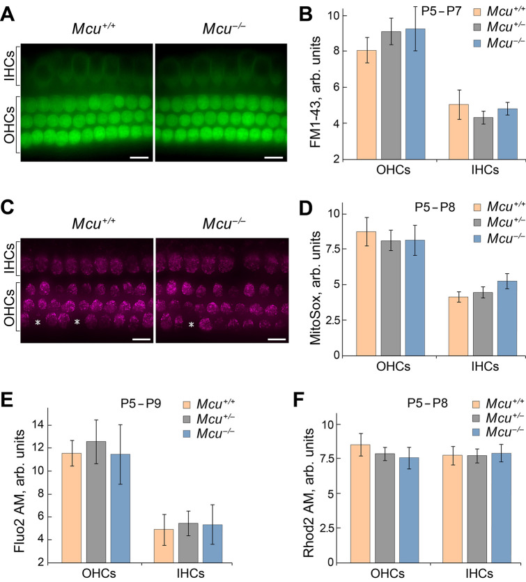
Mcu is not required for the development of hair cells. Results obtained in vitro demonstrate that mechanotransduction, Ca2+ levels, and mitochondria superoxide activity are not altered in the cochlear hair cells of Mcu−/− mice. (A) Representative images show FM1-43 uptake in hair cells of Mcu+/+, Mcu+/− or Mcu−/− mice (mid-basal turn). (B) The fluorescence intensity of FM1-43 in hair cells is not significantly different between the groups, indicating the normal functioning of MET channels in Mcu−/− mice. (C) Representative images showing the MitoSOX indicator levels in hair cells of Mcu+/+or Mcu−/−. Asterisks show Mcu+/+ and Mcu−/− OHCs, which were excluded from statistical analysis due to no detectable superoxide activity. (D) The fluorescence intensity of MitoSOX in hair cells of Mcu−/− mice is similar to that of the Mcu+/+ mice indicating that there is no difference in oxidative activity caused by cellular superoxide buildup. (E,F) The fluorescence intensity of intracellular (Fluo-2 AM) and mitochondrial (Rhod-2 AM) Ca2+ indicators in hair cells of Mcu+/+, Mcu+/− and Mcu−/− mice were not significantly different. Data represented herein are from mice of age P5–P9. Two-way ANOVA analysis did not show any statistically significant differences between Mcu+/+, Mcu+/− or Mcu−/− groups. The number of mice for all groups was greater than 5. Scale bars in (A) and (C) are 12 µm.

