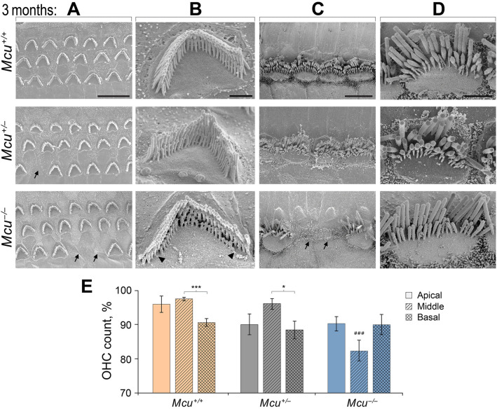
Figure 7.
Loss of hair cells in adult Mcu−/− mouse cochlea. (A–D) Representative scanning electron micrographs of cochlear hair cells acquired from Mcu+/+ (top row), Mcu+/− (middle row), and Mcu−/− FVB/NJ mice (bottom panel) at P88–P92 time point. (A) The typical three-row arrangement of OHCs, (B) single OHC stereocilia at high magnification, (C) the single row of IHCs, and (D) single IHC stereocilia at high magnification are shown. Note the absence of the third (shortest) row of stereocilia in Mcu−/− mice. The arrows in (A) and (C) indicate missing sensory hair cells and the arrowheads in B indicate the degenerating shortest row of stereocilia in OHCs. (E) The SEM images were categorized based on their cochlear region (apical, middle, and basal turn), and the percentage of missing OHCs were plotted (mean ± SEM). ###p < 0.001 in comparison to the middle region of Mcu+/+ and Mcu+/− mice, *p < 0.05, ***p < 0.001, Kruskal–Wallis with Dunn’s multiple comparison tests. The number of mice is 12 (Mcu+/+), 13 (Mcu+/−), and 18 (Mcu−/−). Scale bars are (in µm): 10 (A); 1 (B); 5 (C); 2 (D).

