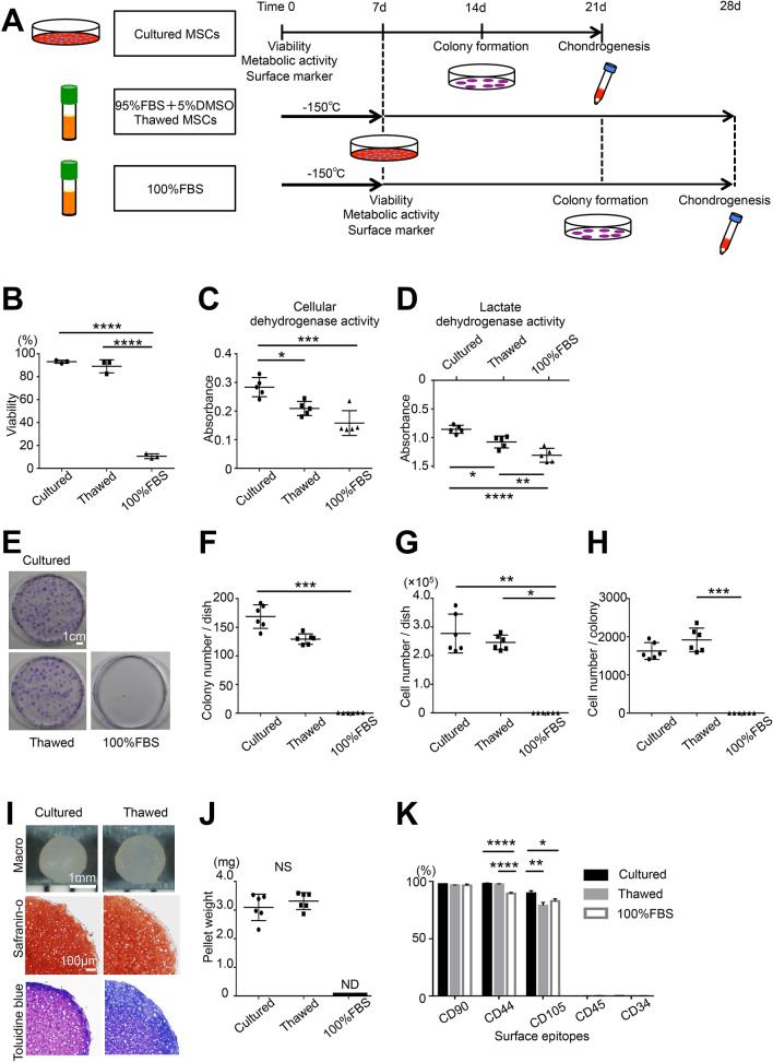
Figure 1.
Effects of cryopreservation on the in vitro viability and properties of rat synovial MSCs. (A) Scheme: 1 × 10⁶ synovial MSCs were suspended in 1000 μL PBS as cultured MSCs. A sample of 1 × 10⁶ synovial MSCs suspended in 1000 μL preservation fluid containing 95% FBS with 5% DMSO or 100% FBS was cryopreserved at − 150 °C for 7 days for use as thawed MSCs or negative controls. The cells were analyzed for viability, metabolic activity, and surface markers. A 0.5 μL volume of cell suspension (containing 500 cells, including living and dead cells) was allocated to a 60 cm2 dish and cultured for colony formation. A 250 μL volume of cell suspension (containing 2.5 × 10⁵ cells, including living and dead cells) was allocated to a 15 mL tube and cultured for chondrogenesis. (B) Viability assessed by trypan blue staining. The average with SD is shown (n = 3). (C) Cellular dehydrogenase activity was used to confirm live cell metabolic activity (n = 5). (D) Lactate dehydrogenase activity was used as a marker of dead cells (n = 5). (E) Colony formation: colonies were stained with crystal violet. (F) Colony number per dish (n = 6). (G) Cell number per dish (n = 6). (H) Cell number per colony (n = 6). (I) Cartilage pellets and histological images. (J) Cartilage pellet weight (n = 6). (K) Surface epitopes (n = 3). ND, not detected; NS not significant; *p < 0.05, **p < 0.01, ***p < 0.001, ****p < 0.0001 by repeated measures one-way ANOVA followed by Tukey’s multiple comparisons (B–D,K), Kruskal–Wallis test followed by Dunn’s multiple comparisons (F–H) or Student’s t-test between two unpaired groups (J).

