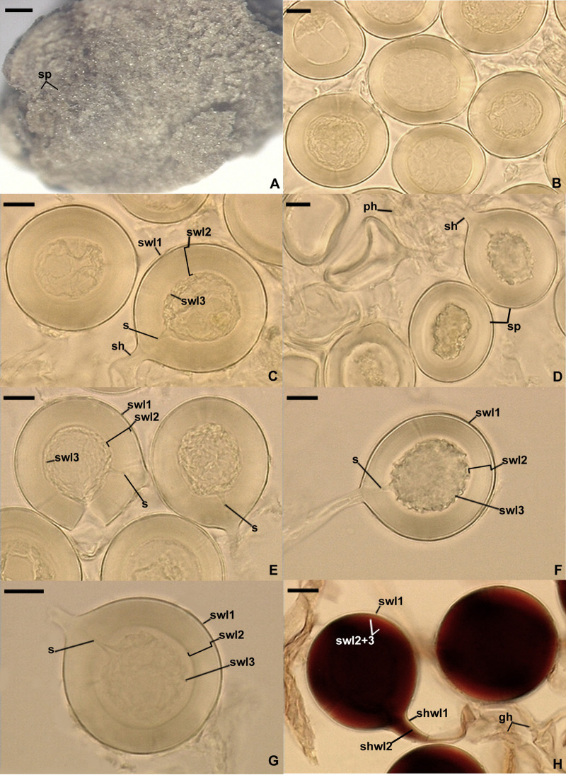
FIGURE 4.
Dominikia glomerocarpica. (A) Glomerocarp (=sporocarp) with glomerospores (=spores; sp). (B) Intact spores. (C–G) Intact and crushed spores with a three-layered spore wall (swl1–3) and a subtending hypha (sh); note that swl2 did not change in shape in crushed spores, and swl3 usually slightly contracted and separated from the lower surface of the laminate swl2 in intact and crushed spores and formed a septum (s) located at approximately half the length of the channel connecting the subtending hyphal lumen with the spore interior; peridial hyphae (ph) are visible in (D). (H) Intact spores with swl1–3, subtending hyphal wall layers (shwl) 1 and 2, and glebal hyphae (gh) mounted in PVLG + Melzer’s reagent. (A) Dry specimen. (B–G) Spores in PVLG. (H) Spores in PVLG + Melzer’s reagent. (A) Light microscopy. (B–H) Differential interference microscopy. Scale bars: (A) = 200 μm, (B–H) = 10 μm.

