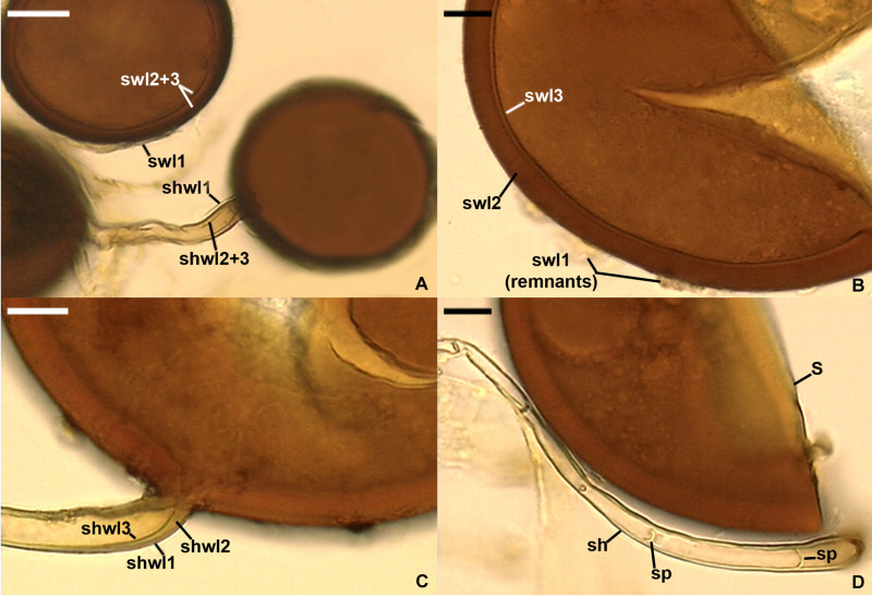
FIGURE 6.
Silvaspora neocaledonica. (A) Spores with a spore wall (sw) consisting of three layers (swl1–3). (B) Spore wall layers (swl) 1–3; swl1 is almost completely sloughed off. (C) Subtending hyphal wall layers (shwl) 1–3. (D) Spore (s) and subtending hypha (sh) with two septa (sp) indicated. (A,B,D) Spores in PVLG. (C) Spores in PVLG + Melzer’s reagent. (A–D) Differential interference microscopy. Scale bars: (A) = 20 μm, (B–D) = 10 μm.

