Figure 1.
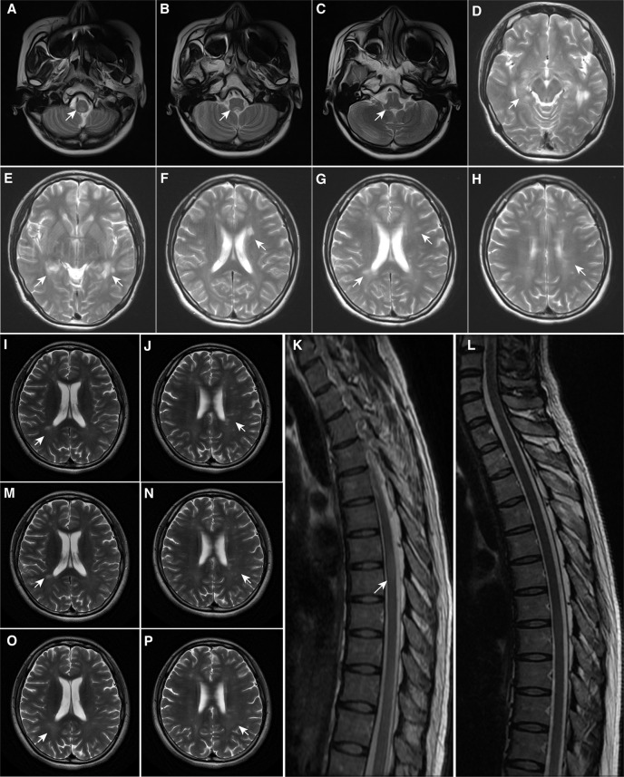
Magnetic Resonance Imaging Findings. (A–C) At first onset, MRI demonstrated patchy lesions in the medulla oblongata and dorsal pons; (D–F) At first recurrence, MRI demonstrated multiple lesions in the brain; (G, H) At second onset, MRI demonstrated pons lesions, consistent with the diagnosis of multiple sclerosis; (I–K) Before teriflunomide administration, MRI demonstrated new punctate enhancing lesions in the left frontal lobe and abnormal signals in the thoracic spinal cord at T6-7 of the skull and spinal cord; (L–N) After 6 months of teriflunomide use, MRI demonstrated that the lesions were reduced, especially in the spinal cord; (O, P) After 1 year of teriflunomide use, MRI demonstrated that the patient’s lesions were reduced. The arrowhead showing lesions in MRI. MRI, Magnetic resonance imaging.

