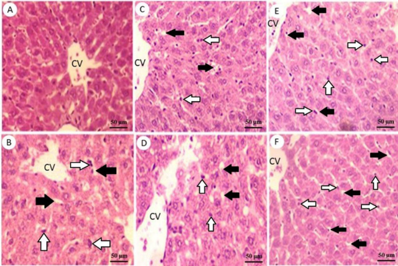
Fig. 2.

Liver sections stained with hematoxylin and eosin. (A) Normal histological view of the liver in control group rats; (B) microscopic examination of liver tissue of untreated diabetic group showing some pathological changes and injury including fat deposit and nuclear pyknosis, which displaying degenerative changes compared to the control normal rats; (C) liver section from an animal treated with glibenclamide (0.25 mg/kg) showing a significant reduction in nuclear pyknosis and fat deposit; (D-F) liver of animals treated with 100, 200, and 400 mg/kg doses of Eryngium billardieri root extract, respectively which indicating the improvement of pathological injury. Magnification: 200⨯. Black arrows, fat deposit; white arrows, nuclear pyknosis.
