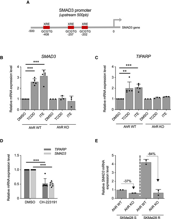
Figure 6. The Transcription Factor AhR Drives SMAD3 Expression.

- AhR binding sites (xenobiotic responsive element (XRE); GCGTG) in human SMAD3 proximal promoter.
- AhR activation by exogenous and endogenous ligands promotes SMAD3 induction. 501Mel cells AhR wild‐type or knockout have been exposed to exogenous and endogenous AhR ligands; TCDD (5 nM) or ITE (10 µM) or the solvent (DMSO) during 10 days. n = 5 biologically independent experiments for AhR WT cells and n = 3 for AhR KO cells. Each histogram represents the mean ± s.d.; multiple comparisons have been done using ordinary one‐way ANOVA, ***P < 0.001.
- AhR activation by exogenous and endogenous AhR ligands promotes TIPARP induction. 501Mel cells have been treated as described in B. n = 5 biologically independent experiments for AhR WT cells and n = 3 for AhR KO cells. Each histogram represents the mean ± s.d.; multiple comparisons have been done using ordinary one‐way ANOVA, **P < 0.01, ***P < 0.001.
- AhR antagonist (CH‐223191) reduces SMAD3 and TIPARP expression levels. SKMel28 cells (AhR wild type) have been exposed to CH‐223191 (5 µM) or the solvent (DMSO) during 7 days. n = 6 biologically independent experiments. Each histogram represents the mean ± s.d.; Bilateral Student test (with non‐equivalent variances): ***P < 0.001.
- Loss of AhR reduces SMAD3 expression levels. SMAD3 expression has been investigated in SKMel28 cells AhR wild‐type (WT) or knockout (KO). SKMel28R has been obtained from SKMel28S by chronic exposure to non‐lethal doses of BRAFi (Hugo et al, 2015b). R for BRAFi‐resistant SKMel28 cells and S for sensitive. n = 2 biologically independent experiments. Each histogram represents the mean ± s.d.
Source data are available online for this figure.
