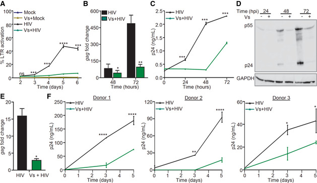
Figure 6. Vs reduces replication of HIV‐1.

-
ACEM‐GFP cells were pre‐treated with 50 ng/μl of Vs for 15 min and infected with 0.1 moi of CXCR4‐using HIV‐1 (NL‐4.3), and GFP fluorescence was measured at 488 nm as an indicator of HIV LTR activity. Vs treatment was repeated every 24 h for the experiment.
-
B–DA similar assay was performed using Jurkat (CD4+ T‐cell line), and viral replication was assessed by (B) gag RT–PCR, (C) p24 ELISA in the culture supernatant, and (D) immunoblotting for p24 (viral capsid protein) in the whole cell lysate.
-
EU937 (promonocytes) were pre‐treated with 50 ng/μl of Vs for 15 min followed by infection with 1 moi of CCR5 using HIV‐1 (NL‐AD8), and viral replication was measured by gag RT–qPCR at 24 h post‐infection (hpi).
-
FPrimary CD4+ T cells purified from human PBMCs (3 healthy donors) were activated, pre‐treated with 25 ng/μl Vs for 15 min, and infected with 0.05 moi of HIV‐1 NL‐4.3. Virus released in supernatant was quantified by p24 ELISA. Vs treatment was repeated every 48 h.
Data information: All figures except (B) and (E) were analyzed by 2‐way ANOVA. (B), and (E) were analyzed by Mann–Whitney test. ****P < 0.0001, ***P < 0.001, **P < 0.01, *P < 0.05. Data are representative of results from three independent experiments performed in triplicate (mean ± SD).
Source data are available online for this figure.
