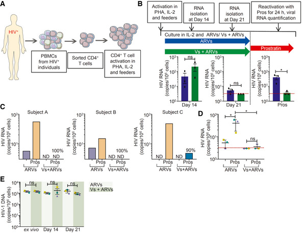
Figure 7. Vs reduces HIV reactivation in primary CD4+ T cells isolated from virally suppressed individuals.

- Schematic representation of generation of expanded CD4+ T cells and reactivation. CD4+ T cells were sorted from PBMCs of ARV‐suppressed HIV‐infected individuals and expanded in presence of PHA, IL‐2, and autologous feeder PBMCs from healthy donor.
- Expanded CD4+ T cells from three patients were cultured in presence of IL‐2 and ARVs, with and without 25 ng/µl Vs for 21 days. Vs treatment was given for 15 min every 3rd day. HIV transcripts were quantified by RT–qPCR at day 14, day 21, and at 24 h post‐stimulation of cells cultured for 21 days by prostratin. Limit of detection for RT–qPCR was 3 viral transcripts per million cells.
- At day 21, cells were stimulated with 1 µM prostratin for 24 h and HIV transcripts were quantified by RT–qPCR. Reduction in viral stimulation in Vs‐treated samples are represented as percentage values. ND—non‐determined.
- Aggregate plot for 3 patients from data (C).
- Total HIV‐1 DNA was determined up to 21 days in cells treated with ARVs or Vs + ARVs.
Data Information: (B), (D), and (E) were analyzed by one‐way ANOVA with Tukey’s multiple correction. *P < 0.05, ns—non‐significant. Data are aggregated from three ARV‐suppressed HIV‐infected human subjects (mean ± SD).
