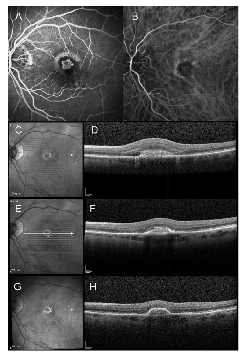
Figure 2.
Spectral domain optical coherence tomography (SD-OCT) findings of an 83-year-old female patient with wet age-related macular degeneration (AMD) and initial type 2 macular neovascularization (MNV). The SD-OCT scans show the progression from type 2 MNV to fibrovascular PED and its margins (at baseline, 1 month after the first intravitreal anti-VEGF injection, and at 1-year follow-up). (A): Early-phase fluorescein angiogram (23 s) at baseline showing a well-demarcated early hyperfluorescence surrounded by a hypofluorescent rim. (B): Early-phase indocyanine green angiogram (21 s) at baseline showing the neovascular network surrounded by hypocyanescent margins. (C): Infrared reflectance photography at baseline, showing the initial hyperreflective ring. (D): Corresponding SD-OCT scan, showing the initial type 2 MNV, with subretinal fluid and hyperreflective exudation. The hyperreflective ring is within the limits of the neovascular lesion. (E): Infrared reflectance photography at 1 month after the first anti-VEGF injection, showing that the hyperreflective ring appeared well-demarcated and retractile. (F): Corresponding SD-OCT scan: the fibrovascular PED can be observed. (G): Infrared reflectance photography at 1-year follow-up: the hyperreflective ring is smaller and has a retractile aspect. (H): SD-OCT scan at 1-year follow-up: the hyperreflective ring corresponds to the limits of the fibrovascular PED lesion.

