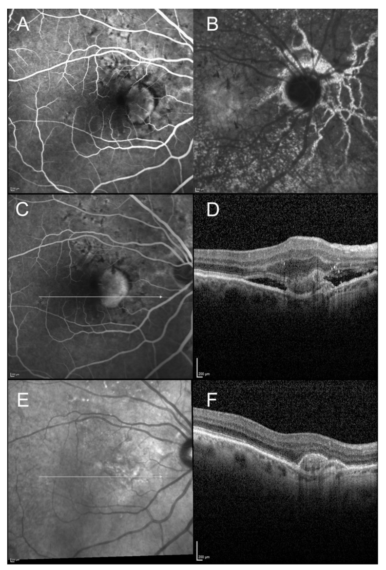
Figure 4.
Imaging of a 64-year-old woman diagnosed with angioid streaks, presenting with visual acuity loss. (A): Fluorescein angiography (22 s) showing the well demarcated hyperfluorescent type 2 neovascular lesion. (B): Indocyanine green angiography (late phase): visualization of the type 2 neovascular lesion is less obvious, but the late phase shows the angioid streaks network clearly. (C,D): Fluorescein angiography and horizontal scan of the spectral domain optical coherence tomography (SD-OCT) at baseline. Well demarcated hyperfluorescent type 2 neovascular lesion, with subretinal fluid and hyperreflective exudation observed on SD-OCT. (E,F): Infrared reflectance photography and horizontal scan of the SD-OCT, at 1-year follow-up. SD-OCT scan shows the fibrovascular PED without any exudation signs.

