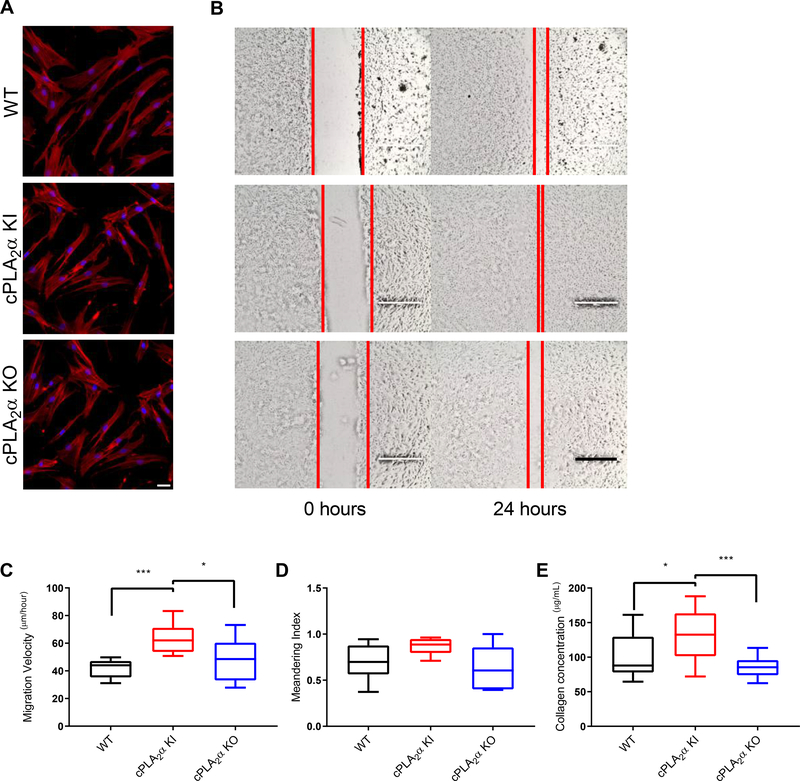
Fig. 5. Loss of the C1P-cPLA2α interaction causes an increase in migration velocity and collagen deposition in primary dermal fibroblasts.
(A) Actin distribution in primary dermal fibroblasts (pDFs) collected from WT, cPLA2α KI, and cPLA2α KO mice. Scale bar, 40μm. (B) Monolayers of pDFs from WT, KI, and KO mice were scratch-wounded and followed for 24 hours. Red lines indicate the borders of the scratch wound. Scale bar, 200μm. (C) Migration velocity of pDFs from WT, KI, and KO mice after scratch-wounding. (D) Quantification of cell meandering in scratch-wounded pDFs from WT, KI, and KO mice. (E) Collagen deposition of pDFs from WT, KI, and KO mice. Samples were analyzed for type I collagen levels using an ELISA-based assay for soluble type I collagen. Data were compared using ANOVA followed by Tukey’s post-hoc test. Data shown are means ± SD, n = 6–12 cell isolates per genotype (5–6 mice per genotype were utilized to generate the cell isolates) for migration and meandering assays, n = 15–16 cell isolates per genotype (8–10 mice per genotype were used to generate the cell isolates) for collagen deposition assay, *P< 0.05, **P< 0.01, ***P< 0.001, ****P< 0.0001.

