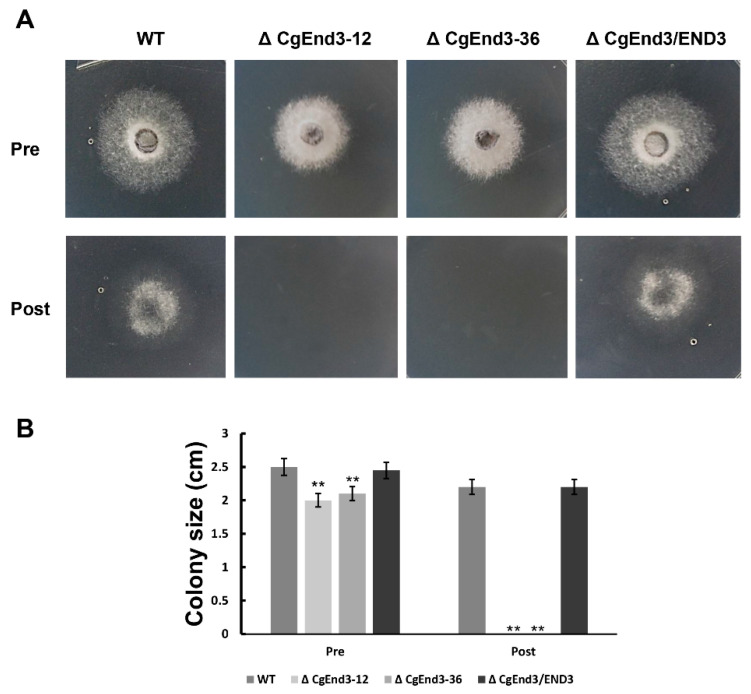
Figure 3.
Cellophane membrane penetration assay. (A) Hyphal blocks from WT, ΔCgEnd3, and ΔCgEnd3/END3 were inoculated on cellophane membranes overlaid on PDA medium for 2 days at 25 °C (Pre). The cellophane membrane were removed, and the resulting plates were incubated at 25 °C for 2 additional days (Post). This experiment was repeated three times. (B) Bar chart showing the colony size of each strain at 2 days post removal of cellophane membrane (Post). Error bars represent the standard deviations. Data were analyzed using Duncan’s range test. Asterisks ** indicate statistically significant differences at p < 0.05.

