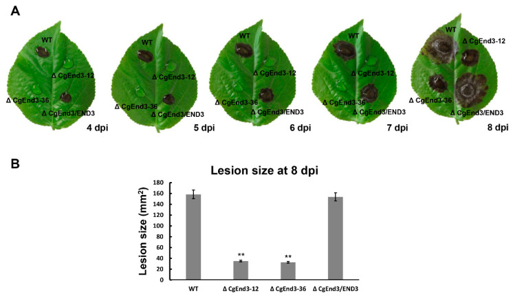
Figure 6.
Pathogenicity assay on poplar leaves. (A) Equal volumes (30 μL) of conidial suspensions (2 × 10⁵ conidia/mL) from WT, ΔCgEnd3, and ΔCgEnd3/END3 were inoculated on poplar leaves. Then, leaves were cultured at 25 °C under moist environment. Images were pictured at 4–8 dpi. This experiment was repeated three times. (B) Bar chart showing the lesion sizes of WT, ΔCgEnd3, and ΔCgEnd3/END3 at 8 dpi. Error bars represent the standard deviations. Data were analyzed using Duncan’s range test. Asterisks ** indicate statistically significant differences at p < 0.05.

