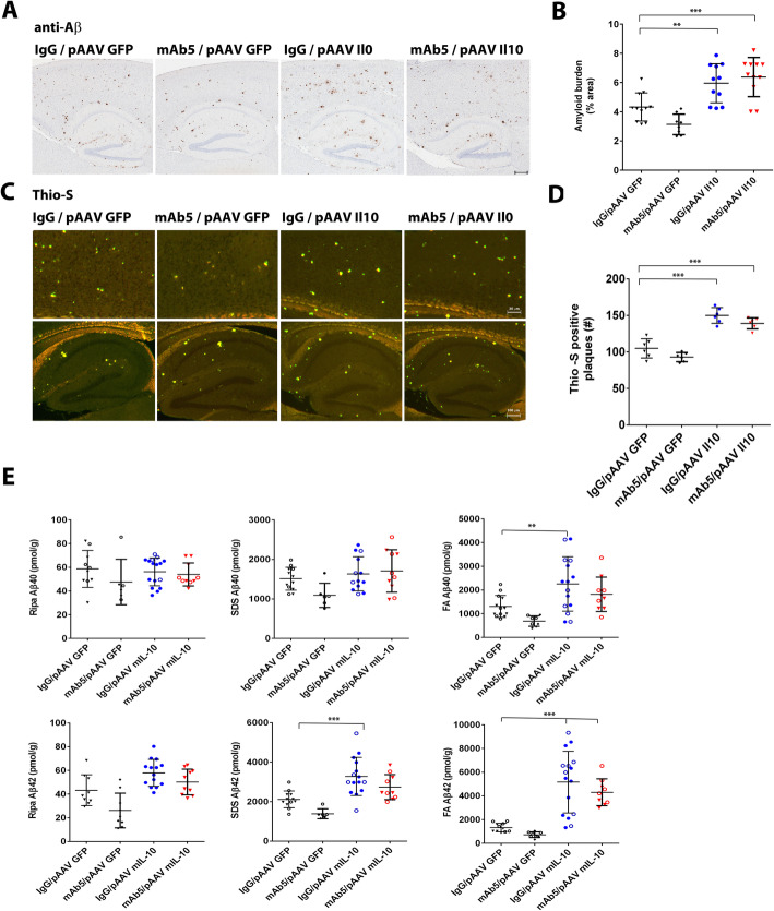
Fig. 4.
Effects of Il10 and mAb5 alone, or in combination on Aβ deposition. a Il10 abolishes the effect of immunotherapy on amyloid loads. Representative brain sections stained with pan-Aβ1–16 antibody (mAb 33.1.1) show Aβ plaque immunoreactivity in the cortex and hippocampus of 6-month-old TgCRND8 mice expressing Il10 or EGFP and immunized with mAb5 or mouse IgG. Scale bar = 150 μm. b The immunostaining was quantified from three sections from each mouse brain using Aperio imaging algorithms. Combination of Il10 overexpression and immunotherapy resulted in increased plaque formation. n = 5–16, *P < 0.05; **P < 0.01. c Overexpression causes an increase in number of Thio-S positive plaques. Il10 overexpression abolished the effects of mAb5 immunotherapy on reduction in the number of Thio-S positive plaques. d Representative fluorescent cortical and hippocampal sections stained Thio-S show Aβ plaque immunoreactivity in the cortex and hippocampus of 6-month-old TgCRND8 mice expressing Il10 or EGFP and immunized with mAb5 or mouse IgG. Scale bars = 150 μm. B. The number of Thio S positive cored plaques was quantified from three sections from each mouse brain. e Il10 overexpression-induced increase in Aβ levels is observed in in cohorts immunized with either mAb5 or mouse IgG as demonstrated by sandwich ELISA with anti Aβ40 and 42 specific antibodies 2.13 and 13.1.1 as capture and 4G8-HRP as detection. Immunotherapy had no effect on Aβ levels in Il10 overexpressing mice. n = 5–16, *p < 0.05, **p < 0.01, ***p < 0.001. Empty and full circles represent male and female mice, respectively

