Fig. 3.
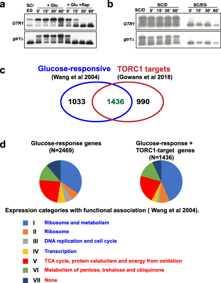
TORC1 activity increases during glucose response. a Wild type and gtr1Δ cells were grown to logarithmic phase in SC/EG medium, and then glucose (2% final concentration) was added to the cultures in the presence of either DMSO or rapamycin (200 nM). Aliquots of the cultures were taken after 0’, 15’, 30’, and 60’ and used for preparing protein extracts. Phosphorylation of Sch9 was monitored by Western blotting. b Wild type and gtr1Δ cells were grown to logarithmic phase in SC/D medium. Cultures were then divided into two parts. For one part, cells were pelleted and washed thrice with SC/EG medium, resuspended in SC/EG medium, and incubated at 30 °C. The second part was transferred back to SC/D and was also incubated at 30 °C. Aliquots of the cultures were taken after 0’, 15’, 30’, and 60’ and used for preparing protein extracts. Phosphorylation of Sch9 was monitored by Western blotting. c Venn diagram showing the overlap of glucose-responsive genes [4] with TORC1 target genes [9]. d Pie chart shows the distribution of the glucose-responsive genes and TORC1-glucose co-regulated (TGC) genes among the various gene clusters defined by response to glucose and Ras activation [4]. Functional enrichment among the various clusters induced and repressed by glucose are indicated, in blue and red fonts respectively

