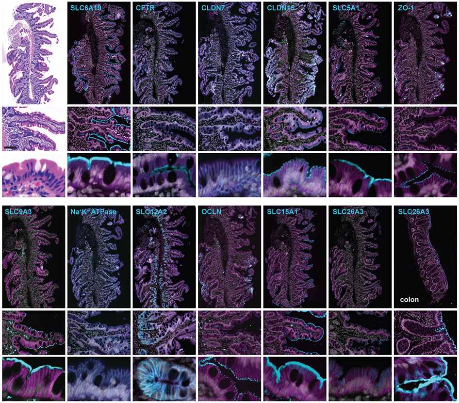
Figure 12. Representative Data.
Serial sections of a duodenal biopsy from a healthy, 26-year-old, female subject were stained by hematoxylin and eosin or with 12 different immunostains. This was one of 50 tissue cores in a single TMA. After imaging and post-acquisition processing using this protocol, pyramidal OME-TIFF images were displayed. Each file (~400 GB) is shown in full (top images, bar = 250 μm), or zoomed in on successively smaller areas (middle images, bar = 100 μm and bottom images, bar = 10 μm). SLC6A19 (Na+-dependent neutral amino acid cotransporter AT1; B0AT1), CFTR (cystic fibrosis transmembrane conductance regulator), CLDN7 (claudin-7), CLDN15 (claudin-15), SLC5A1 (Na+-dependent glucose cotransporter 1; SGLT1), ZO-1 (zonula occludens-1), SLC9A3 (Na+-H+ exchanger 3; NHE3), Na+-K+ ATPase, SLC12A2 (Na+-K+-Cl- transporter 1; NKCC1), OCLN (occludin), and SLC15A1 (H+-peptide cotransporter 1; HPEPT1(Merlin et al., 2001)) are all expressed at appropriate positions along the crypt-villus and basolateral-apical polarity axes. SLC26A3 (downregulated in adenoma; DRA(Gill et al., 2007)), is poorly-expressed in duodenum; but detection and apical expression within surface epithelial are confirmed in proximal colon (bottom right image set). Antibodies used are listed in Table 1.

