FIG 1.
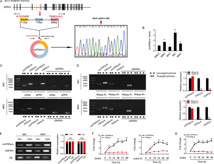
Expression profiles and characterization of circPIKfyve. (A) We confirmed the head-to-tail splicing of circPIKfyve in the circPIKfyve RT-PCR product by Sanger sequencing. (B) Relative expression of circPIKfyve in indicated cell lines was determined by qPCR. (C) RT-PCR validated the existence of circPIKfyve in MIC and MKC cell lines. circPIKfyve was amplified by divergent primers in cDNA but not gDNA. GAPDH was used as a negative control. (D) The expression of circPIKfyve and linear PIKfyve mRNA in both MIC and MKC lines was detected by RT-PCR assay followed by nucleic acid electrophoresis or qPCR assay in the presence or absence of RNase R. (E) circPIKfyve was mainly localized in the cytoplasm, and we used GAPDH as the cytoplasmic marker and U6 snRNA as the nuclear marker. RNA isolated from the nucleus (Nuc) and cytoplasm (Cyto) was used to analyze the expression of circPIKfyve by nucleic acid electrophoresis (up) and qPCR (down); n = 3. (F) qPCR for the abundance of circPIKfyve and linear PIKfyve mRNA in spleen tissues treated with SCRV (MOI, 5) and poly(I·C) at the indicated time points, respectively. (G) qPCR analysis of circPIKfyve and linear PIKfyve mRNA in MKC treated with SCRV (MOI, 5) at the indicated time points. All data represented the means ± SE from three independent triplicate experiments. *, P < 0.05; **, P < 0.01.

