FIG 2.
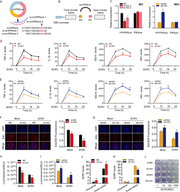
circPIKfyve promotes antiviral innate immunity. (A and B) Schematic diagrams of siRNAs (A) and oe-circ structure (B). (C, left) qPCR analysis of circPIKfyve and linear PIKfyve mRNA in MIC treated with siRNAs. (Right) qPCR analysis of circPIKfyve and linear PIKfyve mRNA in MKC stably overexpressing circPIKfyve. (D and E) qPCR assays were performed to determine the expression levels of TNF-α, IL-1β, and ISG15 or MX1 in MIC transfected with si-circPIKfyve-1 (si-circ) or negative control (NC) (D) and MKC transfected with circPIKfyve overexpression plasmid (oe-circ) or control vector (vector) (E). (F and G) Cell proliferation was assessed by EdU assays in MIC transfected with si-circ or NC (F) and MKC transfected with oe-circ or vector (G). DAPI, 4′,6-diamidino-2-phenylindole. (H and I) Effect of circPIKfyve on cell viability after SCRV infection. MIC and MKC were transfected with si-circ (H) and oe-circ (I) for 48 h and then treated with SCRV for 24 h. Cell viability assays were used. RLU, relative light units. (J and K) circPIKfyve suppresses SCRV replication. MIC and MKC were transfected with NC or si-circ (J) and vector or oe-circ plasmid (K) for 48 h and then infected with SCRV. The qPCR analysis was conducted for intracellular and supernatant SCRV RNA expression. (L) EPC seeded in 48-well plates overnight were treated with cultural supernatants at the dose indicated for 48 h. The cell monolayers then were fixed with 4% paraformaldehyde and stained with 1% crystal violet. si-NC, si-circ, vector, or oe-circ was used. All data represent the means ± SE from three independent triplicate experiments. *, P < 0.05; **, P < 0.01.

