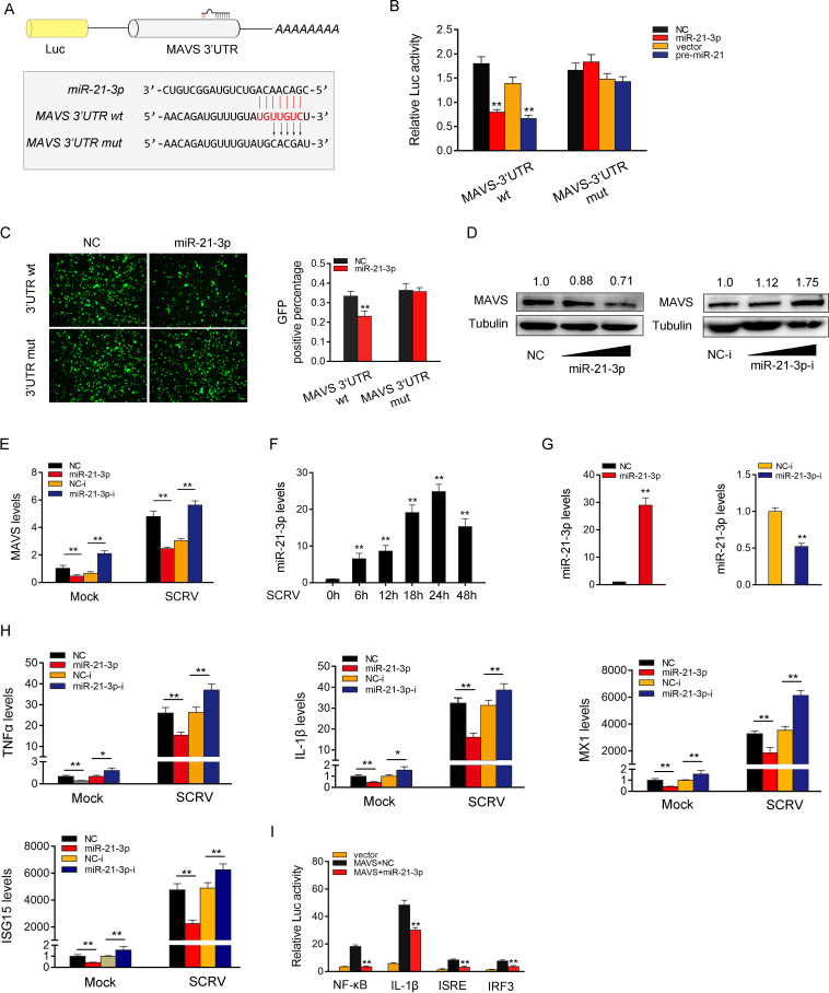
FIG 5.
miR-21-3p inhibits antiviral responses by targeting MAVS. (A) Schematic illustration of MAVS-3′-UTR wt and MAVS-3′UTR mut sequence cloned into luciferase reporter vectors. (B) The relative luciferase activities were detected in EPC after cotransfection with MAVS-3′-UTR wt or MAVS-3′UTR mut and mimics, pre-miR-21 plasmid, or control. (C) EPC were cotransfected with mVenus-MAVS-3′UTR wt or mutated mVenus-MAVS-3′UTR mut, together with NC and miR-21-3p. At 48 h posttransfection, the fluorescence intensity was evaluated by Varioskan LUX (Thermo). (D) Relative protein levels of MAVS were evaluated by Western blotting in MIC after cotransfection with the miR-21-3p mimics or inhibitors. (E) mRNA level of MAVS was evaluated by qPCR under SCRV. (F) The expression of miR-21-3p under different SCRV stimulation times by qPCR. (G) The effect of miR-21-3p mimics and inhibitors on endogenous miR-21-3p expression. MKC were transfected with NC or miR-21-3p (left) and NC-i and miR-21-3p-i (right) for 48 h, and then miR-21-3p expression was determined by qPCR. (H) MKC were transfected with NC, miR-21-3p, NC-i, or miR-21-3p-i. After 48 h posttransfection, the MIC were treated with SCRV for 24 h. The expression levels of TNF-α, IL-1β, Mx1, and ISG15 were analyzed by qPCR. (I) MKC were transfected with NC or miR-21-3p, together with MAVS expression plasmid, phRL-TK Renilla luciferase plasmid, and luciferase reporter genes. The luciferase activity was measured and normalized to Renilla luciferase activity. All data represent the means ± SE from three independent triplicate experiments. *, P < 0.05; **, P < 0.01.

