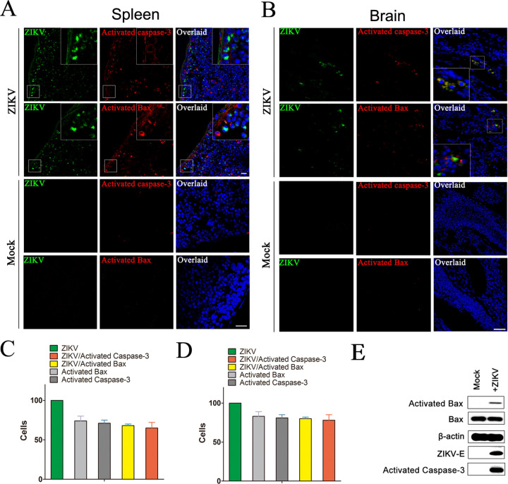
FIG 3.
ZIKV infection is associated with evidence of apoptosis in mice spleen and the fetal brain. (A) Immunofluorescence analysis of the apoptosis in spleen of mice infected with ZIKV. Seven-week-old Ifnar1−/− mice were inoculated with 103 PFU of ZIKV via a subcutaneous route. After 5 days, mice were sacrificed and the spleens were collected for immunofluorescence analysis with ZIKV-E antibody (green), activated caspase-3 antibody (red), and activated Bax antibody (red). DAPI was colored blue. (B) Immunofluorescence analysis of the apoptosis in brain of fetal mice infected with ZIKV. Pregnant Ifnar1−/− mice (6 days post fertilization) were inoculated with 103 PFU of ZIKV via a subcutaneous route. After 7 days, the brain of fetal mice was collected for immunofluorescence analysis with antibody to ZIKV-E, activated Bax, or activated caspase-3. Mock: mice with no ZIKV infection. (C) The ratio of ZIKV positive cells, Bax6A7 positive cells, and both ZIKV and Bax6A7 positive cells in spleen of mice. (D) The ratio of ZIKV positive cells, Bax6A7 positive cells, and both ZIKV and Bax6A7 positive cells in brain of fetal mice. Data are from three independent experiments and the results are presented as mean ± SEM. (E) Western blot of the Bax, activated Bax, and activated caspase-3 in the brain of fetal mice infected with ZIKV. At 7 days postinfection, the brain of fetal mice was collected and homogenized and then immunoblotted with antibody to ZIKV-E, activated Bax, or activated caspase 3. Mock: mice with no ZIKV infection. ZIKV-E: Zika virus’s protein E.

