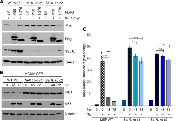
FIG 5.
M50-mediated IRE1 degradation is abolished in SEL1L-deficient cells. (A) WT and Sel1L knockout MEFs were transfected with plasmids expressing Myc-tagged IRE1 and full-length or truncated (1–276) M50 or an empty vector (EV). Cell lysates were harvested at 48 h posttransfection. (B) WT and Sel1L knockout MEFs were infected with MCMV-GFP (MOI = 3). Cell lysates were analyzed by immunoblotting. M57 was detected as an infection control. (C) WT and Sel1L knockout MEFs were MCMV infected as described above for panel B and treated for 5 h with 60 nM thapsigargin (Tg). Total RNA was isolated, and mRNA levels of Xbp1s and Xbp1u were determined by qRT-PCR. Gapdh was used for normalization. Fold changes relative to untreated WT MEFs are shown as means ± SEM from three biological replicates. *, P < 0.05; ***, P < 0.001; ns, not significant.

