FIG 4.
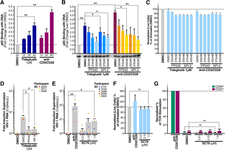
AKT and mTOR modulate tideglusib-mediated NF-κB activation in CD4+ T cells. (A) CD4+ T cells were purified from blood of three HIV-negative individuals and treated for the indicated times with either 1 μΜ tideglusib, anti-CD3/CD28, or a control DMSO solution. (B) Blood CD4+ T cells treated for 1 h with either 1 μΜ tideglusib, anti-CD3/CD28 coated beads, and increasing concentrations of the mTOR inhibitor PP242 or the AKT inhibitor API-2. Nuclear extracts were prepared from these cells (A and B), and p65 binding to target dsDNA was quantitated by enzyme-linked immunosorbent assay (ELISA). Extracts were also immunoblotted for TATA-binding protein (TBP) to test equivalence of protein loading. (C) CellTiter-Blue assays were used to assess potential changes in cell viability in CD4+ T cells treated as for panel B. (D) Fold induction of virion RNA in three HIV-infected individuals on ART treated with tideglusib (1 μM) and DMSO, PP242, or IKK-V, an IKK inhibitor. (E) Fold induction of virion RNA in five HIV-infected individuals on ART treated with SC79 (0.01 to 1 μM) or anti-CD3/CD28-coated beads or left untreated (DMSO control). (F) Normalized percentage of live CD4+ T cells compared to untreated controls. (G) LRA-stimulated CD4+ T cells were tested for surface expression of CD69 and CD25 activation markers. Lymphocytes were gated on single cells > live > CD3+ and CD69+ or CD25+. Unless otherwise specified in the figure, treatments were done at the following concentrations/volumes: DMSO control, 0.01% (vol/vol) final volume; anti-CD3/CD28-coated beads, 125 μl (25 μl per 1 × 10⁶ cells). Data are means and SEM; statistical analysis employed paired two-tailed Student’s t test. *, P < 0.05; **, P < 0.01; ns, not significant.

