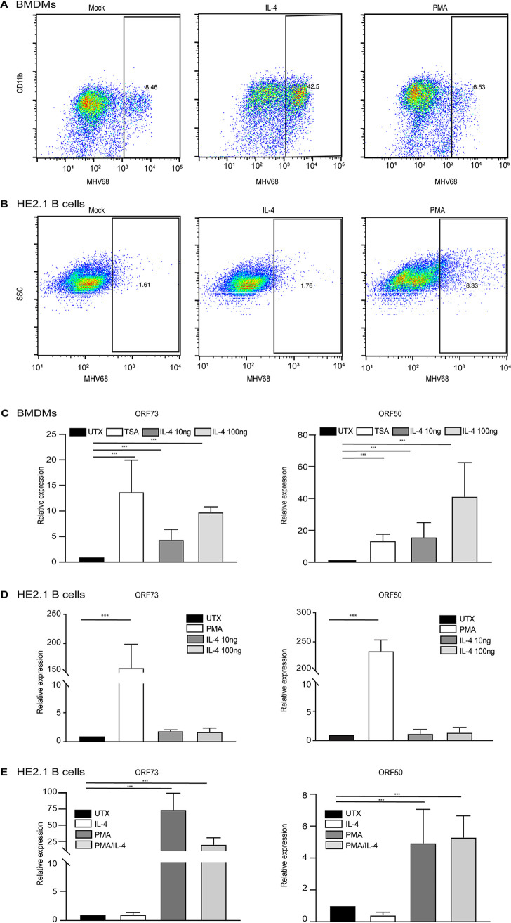
FIG 1.
IL-4 treatment increases viral replication in macrophages but does not reactivate virus from a latently infected B cell line. (A) Bone marrow-derived macrophages (BMDMs) were treated with 100 ng/ml IL-4 and 20 ng/ml PMA for 16 h in culture medium and then infected with MHV68 at an MOI of 5. Twenty-four hours after infection, cells were fixed, and cells expressing lytic viral proteins were determined by flow cytometry. (B) HE2.1 B cells were treated with 100 ng/ml IL-4 and 20 ng/ml PMA for 38 h, cells were then fixed, and cells expressing lytic viral proteins were determined by flow cytometry. SSC, side scatter. (C) BMDMs were treated with 130 nM TSA or different concentrations of IL-4 for 16 h and then infected with MHV68 at an MOI of 5. Transcripts of the virus genes Orf50 and Orf73 were determined 12 h after infection. Expression was normalized to the expression of the glyceraldehyde-3-phosphate dehydrogenase gene (Gapdh). Data are from three independent experiments. (D) HE2.1 B cells were treated with 20 ng/ml PMA or different concentrations of IL-4 for 24 h. RT-qPCR was conducted to assess the expression of the virus genes Orf50 and Orf73. Expression was normalized to the expression of Gapdh. Data are from three independent experiments. (E and F) HE2.1 B cells were treated with 20 ng/ml PMA, 100 ng/ml IL-4, or PMA plus IL-4 for 24 h. RT-qPCR was conducted to assess the expression of the virus genes Orf50 and Orf73. Expression was normalized to the expression of Gapdh. Data are from three independent experiments. UTX, untreated.

