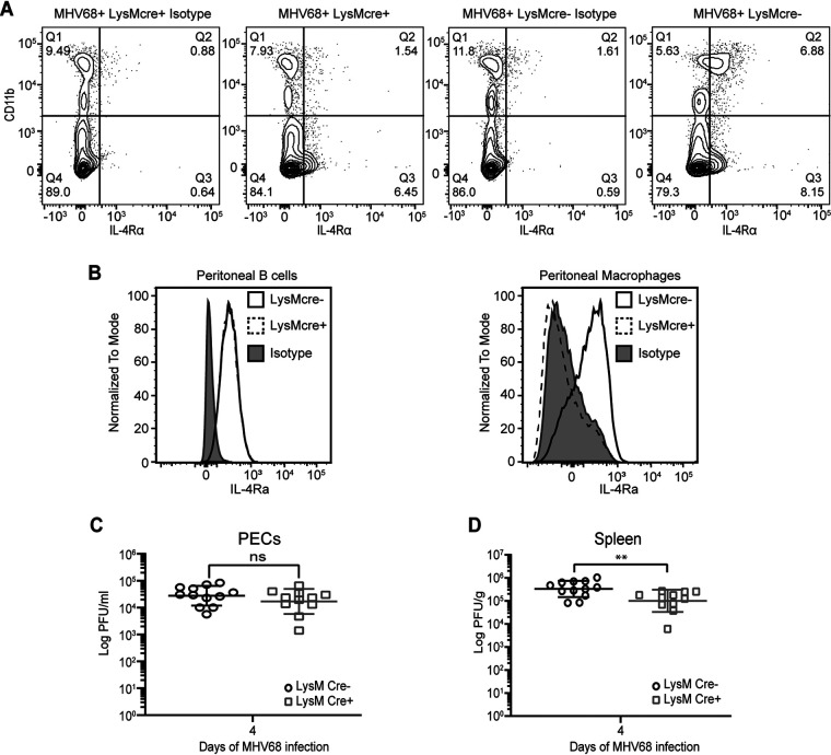
FIG 2.
IL-4R signaling is not required for MHV68 replication. (A) Il4rαf/f × LysMcre+ and Il4rαf/f × LysMcre− mice were intraperitoneally infected with 106 PFU wild-type MHV68. Peritoneal lavage fluid was collected at 37 days post-infection, and flow cytometry was performed to detect IL-4R on peritoneal cells. Cells that have high expression levels of CD11b are macrophages, and CD11b-low or -negative cells include B cells. Contour plots are representative of data from one experiment. (B) Peritoneal exudate cells (PECs) were collected from uninfected Il4rαf/f × LysMcre+ and Il4rαf/f × LysMcre− mice, and flow cytometry was used to detect the IL-4 receptor on peritoneal B cells and macrophages. Representative histograms for two independent experiments are shown. (C and D) Il4rαf/f × LysMcre+ and Il4rαf/f × LysMcre− mice were infected by intraperitoneal injection with 106 PFU of MHV68. PECs (C) and whole spleens (D) were collected at day 4 post-infection, and the virus titer was determined by a plaque assay. Bars represent means ± standard deviations (SD). Each dot represents an individual mouse. ns, not significant; **, P < 0.01 (by a t test).

