FIG 3.
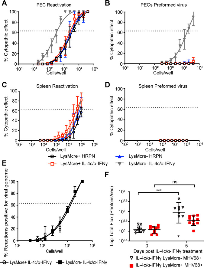
IL-4R expression is required for MHV68 reactivation from macrophages. (A to D) Il4rαf/f × LysMcre+ and Il4rαf/f × LysMcre− mice were intraperitoneally infected with 106 PFU wild-type MHV68. Mice received the isotype control (HRPN) or both IL-4c and anti-IFN-γ at 28 days post-infection. Another dose of IL-4c was administered 2 days after the first treatment, and all mice were then euthanized at 5 days post-treatment. Peritoneal exudate cells (PECs) (A and B) and splenocytes (C and D) were processed into single-cell suspensions. (A and C) Reactivation frequencies were determined by ex vivo plating of serially diluted PECs and splenocytes on a MEF monolayer. (B and D) The presence of preformed virus in PECs and splenocytes was determined by disrupting the cell suspensions and plating serially diluted samples on MEF monolayers. Cytopathic effect was scored at 3 weeks post-plating. Groups of 3 to 5 mice were pooled for each experiment. Data represent the averages from 3 independent experiments. The error bars represent standard errors of the means. (E) Peritoneal exudate cells were processed into single-cell suspensions and subjected to nested PCR analysis to assess the frequency of cells harboring viral genomes. Groups of 3 to 5 mice were pooled for each infection and analysis. The data are pooled from 3 independent experiments. (F) Il4rαf/f × LysMcre+ and Il4rαf/f × LysMcre− mice were intraperitoneally infected with 106 PFU MHV68-M3FL. Mice received anti-IFN-γ and IL-4c after 40 days of infection. IL-4c was administered 2 days after the first treatment, and total flux (photons per second) from the abdominal region was then quantitated at day 5 post-treatment using an Ivis bioluminescence imager. Bars represent means ± SD. Each dot represents an individual mouse. The data are pooled from two independent experiments. ns, not significant; ***, P < 0.001.

