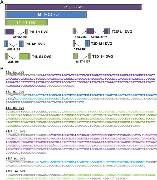
FIG 6.
Schematics of reovirus DVGs. RT-PCR products amplified using primers that bind the 5′ and 3′ termini of the L1, M1, and S4 reovirus segments, smaller than the full-length segments, and indicated in Fig. 2 were excised from agarose gels and sequenced. (A) Schematics, drawn to scale, of reovirus L1, M1, and S4 segments and sequenced rsT1L and rsT3DI reovirus DVGs are shown. The ORF is colored purple (L1), blue (M1), or green (S4), and the 5′ and 3′ UTRs are colored gray. Deletions are indicated by dashed lines, and deleted nucleotides are described. (B) Sequences of sequenced rsT1L and rsT3DI L1, M1, and S4 DVGs. 5′ and 3′ UTRs are colored gray, recombination sites are indicated by a black forward slash, and ORF sequences upstream or downstream of recombination sites are colored in shades of purple (L1), blue (M1), or green (S4).

