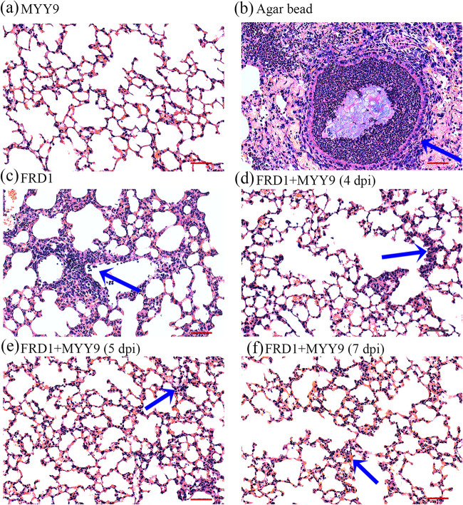
FIG 8.
Histopathological analyses (H&E staining) of inflammatory responses and lung injury following infection with FRD1. (a) The MYY9 group, which was challenged with the therapeutic dose of phage MYY9 alone and no bacterial infection, exhibited no differences compared with normal controls. (b) P. aeruginosa FRD1-laden agar beads colonized the respiratory tract, leading to massive inflammatory cell responses (blue arrow). (c) Phage-untreated group 3 days after infection by FRD1 (3 dpi). (d to f) Lung tissues from the phage-treated group at different time points: 4 dpi (after 24 h of phage therapy) (d), 5 dpi (after 48 h of therapy) (e), as well as 7 dpi (after 96 h of therapy) (f). Compared to the untreated group, the treatment group shown greatly attenuated inflammatory cell infiltration in the perivascular and peribronchial areas. All photos were taken at a magnification of ×200 by an optical microscope. Bars, 50 μm.

