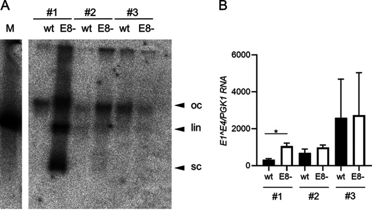
FIG 4.

Analysis of MmuPV1 wt or E8−-positive mouse tail keratinocyte lines. (A) Southern blot analysis of low-molecular-weight DNA isolated from low-passage-number cell lines. DNA was digested with XhoI, a single cutter of MmuPV1 (left), or with EcoRI, a noncutter of MmuPV1 (right). Open circle (oc), linearized (lin), and supercoiled forms of MmuPV1 DNA are indicated by arrows. Linearized MmuPV1 DNA (100 pg) served as a size marker (M). (B) E1^E4 transcript levels were determined by qPCR in total RNA from wt or E8−-positive cell lines, using PGK1 as a reference gene. Average values are derived from three biological replicates, and error bars represent the SEM. Statistical significance was determined by a paired, two-tailed t test (*, P < 0.05).
