Fig. 2.
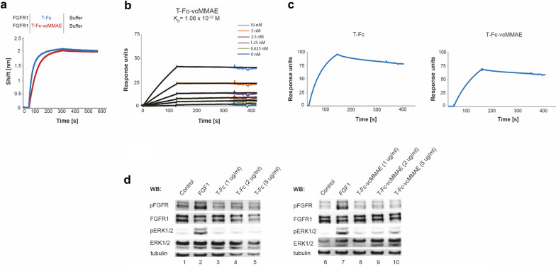
Interaction of T-Fc-vcMMAE with FGFR1. a Evaluation of T-Fc and T-Fc-vcMMAE interaction with FGFR1 by BLI. The extracellular region of FGFR1 was immobilized on BLI sensors and incubated either with T-Fc or T-Fc-vcMMAE. The association and dissociation profiles were measured. b SPR-determined kinetic parameters of the interaction between T-Fc-vcMMAE and FGFR1. The extracellular region of FGFR1 was immobilized on SPR sensors and incubated with various concentrations of T-Fc-vcMMAE. KD value is presented. c SPR results of the interaction between T-Fc and T-Fc-vcMMAE, and murine FGFR1, respectively. The murine recombinant FGFR1 was immobilized on SPR sensors and incubated with T-Fc or T-Fc-vcMMAE. The association and dissociation profiles were measured. d T-Fc and T-Fc-vcMMAE are unable to activate FGFR1. Serum-starved NIH3T3 cells were incubated with FGF1 (positive control) or with different concentrations of T-Fc or T-Fc-vcMMAE. Cells were lysed and activation of FGFR1, and receptor-downstream signaling was assessed with western blotting (WB). The level of tubulin served as a loading control

