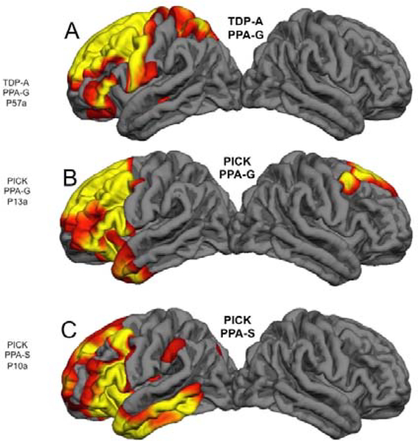
Figure 3: Correspondences of Pathology, Atrophy and Syndrome-.
Quantitative MRI morphometry in 3 right-handed patients who had come to post mortem brain autopsy. Areas of significant cortical thinning compared to controls are shown in red and yellow. A- Onset of PPA-G was at the age of 65. The scan was obtained 2 years after onset. B- Onset of PP-G was at the age of 57. The scan was obtained 5 years after onset. C- Onset of PPA-S was at the age of 62. The scan was obtained 5 years after onset. Despite the differences in neuropathology and clinical syndrome, the one common denominator is the profound leftward asymmetry of atrophy.

