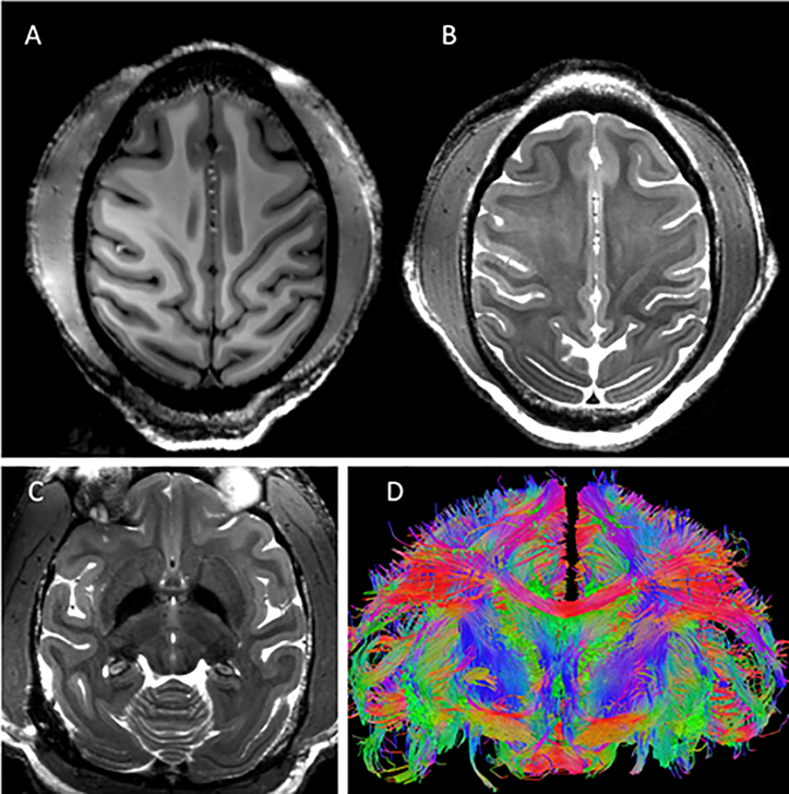
Figure 9:
Representative results obtained with the developed coil. Excellent imaging contrast was achieved at the cortical level with (A) T1 (0.5mm iso: 131×150 mm2 (280×320), TR/TE=3500/3.56ms) and (B and C) T2 (0.4 × 0.4 × 1.0 mm3: 112×150mm2 (288×384),TR/TE =8000/68ms, FA=120°) weighting at 10.5T with a sharp delineation of the white/gray matter borders in superior slices (A & B) which was maintained at deeper brain areas such as the basal ganglia region (C). High-resolution tractography (0.58 mm iso) reconstruction of white-matter bundles in a coronal slice is shown in (D).

