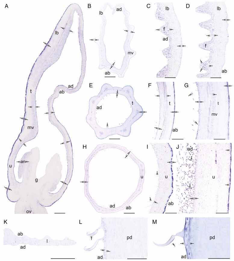
Fig. 5.
In situ hybridization of AfimWIN1. (A) Sagittal section of the Aristolochia fimbriata flower at S6; note higher expression in the adaxial perianth mesophyll, the fimbriae and the abaxial epidermis of the perianth. (B–D) Transverse (B) and longitudinal (C, D) sections of the limb at low (B), mid- (C) and high (D) magnification. (E–G) Transverse (E) and longitudinal (F, G) sections of the tube at low (E), mid- (F) and high (G) magnification. (H–J) Transverse (H) and longitudinal (I–J) sections of the utricle at low (H), mid- (I) and high (J) magnification. (K) Transverse section of the leaf. (L–M) Longitudinal section of the floral pedicel (L) and detail of its hooked trichomes (M). Arrow/bar signs point to tissue with positive expression; ab, abaxial epidermis; ad, adaxial epidermis; an, anther; fi, fimbriae; g, gynostemium; l, leaf; lb, limb; mv, midvein; ov, ovary; pd, pedicel; t, tube; u, utricle. Scale bars: 100 µm (A, B, E, H); 150 µm (C, F, I); 200 µm (D, G, J); 60 µm (K–M).

