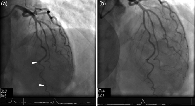
Fig. 1.

(a) A cranial (37°) coronary angiographic view illustrating a typical type-two coronary artery dissection at the distal left anterior descending artery (region between the two arrowheads). (b) Follow-up coronary angiography [projection approximately same to (a)] demonstrating total restoration of the patency of the affected vessel. CRA, cranial; LAO, left anterior oblique.
